Table of Contents
Cover
Website
Title page
Copyright page
Dedication
Preface
Acknowledgments
SECTION 1: ANATOMY
Chapter 1 Directional Terms
Chapter 2 The Common Integument
Introduction
Skin
Pads
Haircoat
Claws
Species differences
Chapter 3 Skeletal Anatomy
Introduction
The long bones
Flat bones
Irregular bones
Equines
Bovines
The bone that isn’t
Bird bones
Chapter 4 Muscle Anatomy
Introduction
Skeletal muscle
The head and neck
The thorax
The dorsum
The abdomen
The pelvis
The limbs
Smooth muscle
Chapter 5 The Anatomy of Joints
Introduction
Types of joints
Tendons
The skull
The ribs and vertebral column
The pelvis and hip
The shoulder and thoracic limb
The pelvic limb
Chapter 6 Anatomy of the Nervous System
Introduction
The neuron
The brain
The spinal cord
Peripheral nervous system
Chapter 7 Anatomy of the Urinary Tract
Introduction
The kidneys
Leaving the kidney
The urinary bladder
Chapter 8 Cardiovascular Anatomy
Introduction
The heart
The exterior of the heart
The interior of the heart
Peripheral circulation
The named arteries
The veins
The lymphatic system
Chapter 9 Anatomy of the Digestive System
Introduction
Canines and felines
The oral cavity
The pharynx and the esophagus
The stomach
The intestines
The liver
Species variation
Chapter 10 Anatomy of the Endocrine System
Introduction
The hypothalamus and the pituitary
The peripheral endocrine system
Chapter 11 Respiratory Anatomy
Introduction
Entry into the respiratory system
The larynx
The trachea and lungs
Species differentiation
Chapter 12 Reproductive Anatomy
Introduction
The female
The male
SECTION 2: PHYSIOLOGY
Chapter 13 The Cell
Introduction
Mammalian cell boundaries
The production of energy
Chapter 14 Functions of the Common Integument
Introduction
Skin
Glands
Hair
The pads
Antlers and horns
Chapter 15 Osteology
Introduction
The growth of bones
Bone marrow
Cartilage
Ligaments, tendons, and joints
Avians
Chapter 16 Muscle Physiology
Introduction
Locomotion
The generation of a muscle contraction
The basis of speed
Smooth muscle
Chapter 17 Sensory Physiology
Introduction
Receptors
The visual system
Proprioception
The auditory system
The olfactory system
The gustatory system
The tactile system
Specialized receptors
Chapter 18 Neurophysiology
Introduction
The neuron
The action potential
Central and peripheral functions
The brain
The autonomic nervous system
Chapter 19 Renal Physiology
Introduction
The nephron
Renal excretion/resorption of water
Acid/base balance
Blood pressure and the renal system
Anemia and the kidney
Species differences
Chapter 20 Cardiovascular Physiology
Introduction
The heartbeat
Cardiac output
Flow through the heart and back
Blood pressure
Autonomic nervous system involvement
The lymphatic system
Chapter 21 Digestive Physiology
Introduction
The entryway
The stomach
Entering the intestine
The liver
Species differentiation
Chapter 22 Endocrine Physiology
Introduction
Endocrine glands
The pituitary
The thyroid
The parathyroid
The adrenal gland
The pancreas and insulin
Other endocrine activities
Chapter 23 Respiratory Physiology
Introduction
The basics
Ventilation and temperature
Residual capacity
Thoracic pressure
The nervous system and respiration
The rhythmicity of breathing
Gas exchange
Hemoglobin
Carbon dioxide
Chemoreceptors
Mechanisms to increase oxygenation
Species differences
Chapter 24 Reproductive Physiology
Introduction
A cascade of events
The female
Hormonal control
Other factors affecting the estrous cycle
The male
Fertilization and pregnancy
Parturition
Appendix 1 Dissection Notes
Appendix 2 The Cranial Nerves
Appendix 3 Selected Muscle Origins and Insertions
From origin to insertion
Answers
Glossary
References
Index
Website
This book is accompanied by a companion website:
www.wiley.com/go/sturtz
The website includes:
- The figures from the book for downloading into PowerPoint
- Additional questions and answers
- Labeling quizzes
- Teaching PowerPoints
- A dissection video
This edition first published 2012 © 2012 by John Wiley & Sons, Inc.
Wiley-Blackwell is an imprint of John Wiley & Sons, formed by the merger of Wiley’s global Scientific, Technical and Medical business with Blackwell Publishing.
Editorial offices: 2121 State Avenue, Ames, Iowa 50014-8300, USA
The Atrium, Southern Gate, Chichester, West Sussex, PO19 8SQ, UK
9600 Garsington Road, Oxford, OX4 2DQ, UK
For details of our global editorial offices, for customer services and for information about how to apply for permission to reuse the copyright material in this book please see our website at www.wiley.com/wiley-blackwell.
Authorization to photocopy items for internal or personal use, or the internal or personal use of specific clients, is granted by Blackwell Publishing, provided that the base fee is paid directly to the Copyright Clearance Center, 222 Rosewood Drive, Danvers, MA 01923. For those organizations that have been granted a photocopy license by CCC, a separate system of payments has been arranged. The fee codes for users of the Transactional Reporting Service are ISBN-13: 978-0-8138-2264-8/2012.
Designations used by companies to distinguish their products are often claimed as trademarks. All brand names and product names used in this book are trade names, service marks, trademarks or registered trademarks of their respective owners. The publisher is not associated with any product or vendor mentioned in this book. This publication is designed to provide accurate and authoritative information in regard to the subject matter covered. It is sold on the understanding that the publisher is not engaged in rendering professional services. If professional advice or other expert assistance is required, the services of a competent professional should be sought.
Library of Congress Cataloging-in-Publication Data
Sturtz, Robin.
Anatomy and physiology for veterinary technicians and nurses : a clinical approach / Robin Sturtz, Lori Asprea.
p. cm.
Includes bibliographical references and index.
ISBN 978-0-8138-2264-8 (pbk. : alk. paper) 1. Veterinary anatomy. 2. Veterinary physiology. I. Asprea, Lori. II. Title.
SF761.S87 2012
636.0892–dc23
2012012163
A catalogue record for this book is available from the British Library.
Wiley also publishes its books in a variety of electronic formats. Some content that appears in print may not be available in electronic books.
Disclaimer
The publisher and the author make no representations or warranties with respect to the accuracy or completeness of the contents of this work and specifically disclaim all warranties, including without limitation warranties of fitness for a particular purpose. No warranty may be created or extended by sales or promotional materials. The advice and strategies contained herein may not be suitable for every situation. This work is sold with the understanding that the publisher is not engaged in rendering legal, accounting, or other professional services. If professional assistance is required, the services of a competent professional person should be sought. Neither the publisher nor the author shall be liable for damages arising herefrom. The fact that an organization or Website is referred to in this work as a citation and/or a potential source of further information does not mean that the author or the publisher endorses the information the organization or Website may provide or recommendations it may make. Further, readers should be aware that Internet Websites listed in this work may have changed or disappeared between when this work was written and when it is read.
Dr. Sturtz would like to thank LVTs Asprea and Sternberg and dedicates this book to her students, teachers, family and pets, which should about cover it.
Lori Asprea (coauthor, photography) thanks her mother and father, sister, and entire family; Mercy College and the Vet Tech Department; and, of course, Robin Sturtz and the animals. Without them, this book would be very boring. I would like to dedicate this book to my family, who has endlessly supported my ambitions.
Robin Sternberg (illustrations) thanks Drs. Jennifer Chaitman, Anthony Pilny, and Eveline Han, and would like to dedicate this book to Dr. Jennifer Chaitman—my mentor, my friend, and my inspiration.
Preface
The authors and the illustrator have all been students as well as teachers of veterinary science. We have long talked about putting together a book that reflects how we think about the subjects of anatomy and physiology. For one thing, we sought to have the anatomy and the physiology portions of the book as two separate sections. In some veterinary technology programs, anatomy and physiology are actually taught as separate courses. Even when they are combined, it can be more helpful to build on the foundation of a complete understanding of the anatomy in order to understand the complexity of physiology.
We all agree that adding clinical scenarios makes the information more interesting and, thus, easier to remember. Anatomy and physiology are not part of our curriculum for the sake of theory. The aim of the study of these topics is to be able to apply this knowledge to daily practice in a clinic, hospital, or research facility.
In this first edition, there are materials that have no doubt been excluded that might have been included, or concepts that were included but do not strike the readers as interesting to the same level that the authors find them to be. We fervently hope that this work will be of use. We hope equally as much that you, the reader, will let us know what information you would like added or subtracted as we move forward. Please note that any errors are solely the responsibility of Dr. Sturtz.
We hope that you find this book worthwhile, not only for current study but also for frequent reference. There are also online components that will be of help for people who learn best with visual images rather than written narrative.
Robin Sturtz
Lori Asprea
Acknowledgments
It is absolutely impossible to write and illustrate a book like this without the support and guidance of dozens of people. It is possible to go on for pages of excruciating detail thanking everyone who has helped us with this project. However, in deference to the reader, we shall endeavor to be brief.
The guidance of our teachers has sustained us and carried us to this moment. We thank the faculty and staff at all of the colleges we have attended (the authors have Mercy College in Dobbs Ferry, New York, in common). We thank our family and friends for their tremendous forbearance. We particularly thank our students for their enthusiasm, their patience, and their encouragement, not to mention their perfect timing when it comes to asking that one last question …
Our good friends at John Wiley & Sons are, of course, the ones who made this all happen. To Nancy and all of the editorial wizards there, our deepest gratitude.
Dr. Sturtz would like to honor a few special people: Professors Buell, Burke, and Burke at Mercy College; Drs. C. Brown, S. Brown, P. Carmichael, and S. Allen at The University of Georgia College of Veterinary Medicine; friends and colleagues at La Guardia Community College, Mercy College, and The Cat Hospital; the veterinary Bregman family; Kim, Ceasar, Judy, Glen, and Jon, and a whole host of others. Kudos to Elaine Sturtz for last-minute artwork. (Yay mom!) If I’ve met you, count yourself in.
And, of course, we all thank our pets, who could probably write this book just as well as we did, but who preferred to sit back and laugh at us while we did it. Good going, pets!
R.S.
L.A.
SECTION 1
ANATOMY
Chapter 1
Directional Terms
Clinical case: Fluffy
Clinical case resolution: Fluffy
Review questions
Clinical Case: Fluffy
A veterinary technician comes in for work in the morning to discover that Fluffy has come in overnight after having been hit by a car. The chart note indicates that there is “a cut on the back leg.” This, of course, gives very little information.
The study of anatomy is, put simply, the study of the structure of organisms. It involves looking at architecture, at the different positions, shapes, and sizes of various living tissues. As one might imagine, the anatomy of different species has some things in common and some things that are quite diverse. The structure of the heart is very similar in dogs and cats; it is quite different in equines and reptiles. The kidneys of the dolphin look very different from those of the dog, although they function in the same way. By understanding the differences in anatomy among animals, we can have a greater appreciation for how their body systems function. This understanding is the basis of recognizing states of health and disease.
There are a number of different ways to organize how one looks at anatomy. Gross anatomy refers to features that can be seen with the naked eye. Developmental anatomy is the study of how anatomy changes as the fetus becomes a puppy or a kitten. Topographic anatomy refers to the relation to the parts to the whole (e.g., how the different parts of the kidneys and the connecting conduits make up the urinary system). Regional anatomy refers to the structures of a given area of the body; if one looks at the head, for example, as one unit, it would involve the study of all the muscles, blood vessels, bones, and other tissues that are present. Imaging anatomy refers to the anatomical features as they are seen on a good radiograph. Applied anatomy refers to the anatomy that is most important surgically or for medical treatment. In planning orthopedic surgery, for instance, it is necessary to know not only the structure of the bones but also the local muscles and blood vessels. Most of us use a systems approach. We study all of the bones, then all of the muscles, then all of the digestive organs, without regard to where they are placed.
One of the most important issues in studying anatomy is the understanding of directional terms. If one is asked to find a particular spot on the animal, having someone say “It’s on the leg” is not terribly precise. Saying that a spot is “just proximal to the right stifle” (just above the right knee) is much clearer. While the acquisition of vocabulary can be tedious, it is the way to communicate clearly with our clients, veterinarians, and other members of the patient-care team. In other words, good anatomic vocabulary contributes to excellent patient care.
Directional terms in veterinary medicine are very different from those used in human medicine. The human head is “up” from the hips, while it is “forward” in the dog. This is another reason that it is important to use the proper terms.
Going toward the head is considered to be cranial; going in the opposite direction is caudal. Going from the floor upward is traveling in a dorsal direction. From the top downward is moving in a ventral direction. On the limbs, we use special terms. Closer to the body is proximal; going away from it is moving in a distal direction (Figure 1.1).
Moving toward the center is going medially. Going out from the midline to the side of the animal is a lateral movement. On the head, there is a special term. When we discuss something that is cranial to another spot on the head, we describe it as rostral (from the Latin word for face).
We need to add some more vocabulary to refer to specific places. The part of the body that includes the chest and abdomen is referred to as the trunk. The proper name for the ventral part of the abdomen is the ventrum. The proper name for the top of the trunk is the dorsum. The lateral surface of the part of the trunk caudal to the chest is the flank.
There are specific names for other parts of the body. The part of the trunk from the neck to the caudal ribs is referred to as the thorax. The areas around the ribs are referred to as costal areas. The abdomen refers both to the outer surface (the skin) of the ventrum and also to the space within it. The thoracic inlet is the area of the ventral thorax where the neck ends.
The space within the thorax is called the thoracic cavity, and the space within the abdomen is the abdominal cavity. Note, however, that some of the features of each of these are lined by a membrane. The pleural membrane, within the thorax, surrounds the lungs and lines the walls of the thoracic cavity. The area bordered by this membrane is considered to be within the pleural cavity. Similarly, the membrane surrounding some of the organs and lining the interior walls of the abdomen is the peritoneal membrane, or peritoneum, and that space is called the peritoneal cavity. Inflammation of that membrane is called peritonitis. There is a viral disease called feline infectious peritonitis that causes inflammation of the peritoneum (among other things). The localized inflammation is so severe that a condition called ascites (fluid in the abdomen) can result. This disease is incurable and can only be treated symptomatically.
Features of the limbs get special names. The front legs are referred to as the thoracic limbs, the rear legs as the pelvic limbs. The shoulder and elbow in dogs are, in medical terms, the scapulohumeral and humeroradioulnar joints, respectively. The next joint distal to the elbow is the carpus, the equivalent of the human wrist. The front of the leg from the shoulder going distally to the paw is the dorsal section, with the back of that area up to the carpus referred to as the caudal section of the limb. On the pelvic limb, the joint between the femur and the tibia is the femorotibial joint, commonly known as the stifle; it is equivalent to the human knee. The next joint going distally is the tarsus. The common name for the tarsus is the hock. The same terms, dorsal and caudal, apply to the pelvic limb.
The part of the thoracic limb from the shoulder to the elbow is referred to as the brachium; the area from the elbow to the carpus is the antebrachium. The area from the head of the femur (the proximal-most bone of the pelvic limb) to the stifle is called the femoral area. The area from the stifle to the tarsus technically is called the crus, although that term is not commonly used in a clinical setting; distal pelvic limb is less precise but often used in general practice.
There is one other directional label referring to the limbs. The area from the carpus distally, on the caudal surface, and around to and including the ventral surface of the paw, is known as the palmar surface. The analogous area from the tarsus to the bottom of the paw is the plantar surface.
The joints of equines have a number of common names that are distinct from those of small animals. For example, the carpus is called the knee, and the tarsus is called the hock.
There are different names for the bones themselves in large animals. The long bone distal to the carpus and tarsus is known as the cannon (equivalent to the third metacarpal/metatarsal bone of dogs and cats). The three bones of the digit, called proximal, middle, and distal phalangeal bones, have a different set of names. They are called, respectively, long pastern, short pastern, and coffin bone. We will discuss the specific names of bones in Chapter 3.
Clinical Case Resolution: Fluffy
In the example at the beginning of this chapter, a problem was noted with Fluffy’s “back leg.” The chart note is not terribly precise. We can record that Fluffy has a laceration proximal to the right stifle, on the medial surface of the limb, and it will be clear to any reader exactly where the problem is. This is the advantage of using correct anatomical terms.
Review Questions
1 Define the terms medial, rostral, and dorsal.
2 Which is more cranial, the thoracic limb or the pelvic limb?
3 The caudal paw area on the thoracic limb is referred to as the _____ surface.
4 True or false: The stifle is caudal to the tail.
5 Define the term “topographic anatomy.”
Chapter 2
The Common Integument
Clinical case: Cat with alopecia
Introduction
Skin
Pads
Haircoat
Claws
Species differences
Clinical case resolution: Cat with alopecia
Review questions
Clinical Case: Cat With Alopecia
A cat is brought into the clinic for a general checkup and vaccines. There is an area of alopecia (missing hair) that is perfectly round. It is located on the dorsal surface of the animal’s head and is about 0.5 cm in diameter. The skin is not irritated, and the client does not report that the cat has been scratching it.
The term integument refers to a broad range of tissues. Knowing the composition and structure of these elements contributes to a broader understanding of the function of this system. This will lead to the ability to recognize what happens in disease states such as the one described above.
Introduction
The integument is a collective term for aspects of bodily structure that are formed of connective tissue and epithelia. Connective tissue is a collection of proteins, fibrous material, and ground elements that form a great many parts of the mammalian body. An epithelial cell has a specific microscopic structure. Features such as skin, skin glands, fur/whiskers, hooves, horns, and claws are epithelial structures that are parts of the integument. This chapter will focus on mammals; certain species-specific characteristics will be discussed at the end of the chapter.
Skin
Mammalian skin has several layers to it. The superficial-most layer is the epidermis. Deep to it is the dermis. There is a layer of fat and connective tissue deep to the dermis called the subcutaneous layer. The subcutaneous layer is not skin (subcutaneous means below the surface of the skin) but is a part of the integument.
The epidermis itself has a number of layers. The deepest of these is the stratum basale. Continuing toward the surface, the layers are the stratum spinosum, stratum granulosum, stratum lucidum, and stratum corneum. See Figure 2.1 for a depiction of these layers.
Some parts of the epidermis have more layers than others. For example, there is no stratum lucidum in areas where there are hair follicles, or where the skin is very thin. In areas where skin is thick, such as the specialized epidermis known as the pads of the paw and limb, there may be a relatively thick stratum lucidum.
The stratum lucidum and stratum corneum (from the Latin root for “horn”) are composed of cells that are, for lack of a better word, dead. When the living cells are formed in the deepest layers of the epidermis, they work their way up toward the surface, dying along the way. By the time they reach the most superficial layer of the epidermis, the stratum corneum, they have lost their nuclei and are ready to slough off. These compose the flakes of “dry skin” that are noted on gross observation.
The epidermis has almost nothing in the way of blood vessels or nerve fibers. This is why a cat’s head can be gently scratched without drawing blood. Anything that penetrates down to the level of the dermis, however, will cause pain and bleeding, as that is where the blood vessels and nerves are located.
The dermis is mostly composed of collagen, a protein. The bases of individual hairs are encased in a space called a follicle; the majority of the follicles lie within the dermis. The dermis is also where the nerves and blood vessels run and where the skin glands are found. It is a layer that has primary importance in the functions of skin as well as in structural support.
Dermal glands fall into two main categories: sweat and sebaceous. Sebaceous glands produce sebum, which is a waxy, thick substance. Sebaceous glands also secrete pheromones, which are species-specific odors that have a great deal to do with socialization and reproductive behaviors. There are a number of sebaceous glands throughout the body, in all mammalian species. The interdigital glands are important in ruminants. These are present in the area of the hoof where the digits begin to spread out from each other. The anal glands, present on either side of the anus, assume an important communication role in dogs and cats and will become quite familiar in clinical practice. Suborbital glands, present in the area of the medial canthus (place where the eyelids meet) of each eye, are used to mark territory by many antelopes.
Sweat glands are of two types, apocrine and eccrine. These types differ in their function and will be discussed in Chapter 14. In general, apocrine sweat glands are found in association with hair follicles, while eccrine glands are spread in other areas. Canines and felines have sweat glands primarily around the metacarpal and digital pads of the paw. A specialized form of sweat gland is the mammary gland.
A discussion of the skin would not be complete without a discussion of the subdermal or subcutaneous layer (usually abbreviated as “sub-Q” or “SQ”). This layer is mostly composed of adipose tissue (i.e., fat). When a cat or a dog picks a neonate up by the back of the neck, it is not painful. This is because she is holding him/her by the scruff. The scruff is the part of the body on the dorsal neck that has a very thin layer of skin, and is mostly composed of subcutaneous fat. Sub-Q, like the epidermis, has little in the way of blood vessels or nerve fibers. In a healthy animal, this layer has a large water content; in a dehydrated animal, areas of thick subcutaneous tissue, such as the scruff, will maintain a tented appearance if pinched and released, rather than springing back to a normal position. This is one way we assess hydration status during a physical examination.
Pads
A thickened mass of epidermal layers is referred to as a pad (see Figure 2.2). Dogs and cats have a pad on the palmar/plantar surface of each digit and a larger pad proximal to it called a metacarpal or metatarsal pad, depending on the limb (carpal always refers to the thoracic limb and tarsal to the pelvic limb). There is also a small pad in cats just proximal to the carpus. This pad is usually associated with one of the skin glands and is often marked by a single tactile hair.
Horses have pads on the limbs, referred to as the chestnut and ergot. The chestnut is found on the medial surface, in the area of the carpus/tarsus. Interestingly, chestnuts are thicker in working horses than in light breeds, where they may be absent altogether. The ergot is on the caudal surface in the same area as the chestnut on each limb. A concentration of sebaceous glands in this area may be involved in scent marking.
Haircoat
The haircoat is an important feature of the integument. One of the attributes that defines an animal as a mammal is that it has hair.
In dogs and cats, the haircoat has several layers. The word topcoat is used to denote the outermost layer. The trapping of air between the coat and the undercoat is an important method of thermoregulation (see Chapter 14 for a further discussion of thermoregulation). The undercoat is usually thicker than the topcoat and is the layer that does most of the shedding. When a dog or cat is combed, it is mostly the undercoat that comes off onto the comb. The topcoat is composed of guard hairs. The hairs of the undercoat are referred to as wool hairs.
The whiskers (vibrissae) are a type of hair called tactile hair. They are thicker than guard hairs and are only present at certain places on the body; in mammals, they tend to cluster on the face. They sense motion and objects and should never be trimmed unless medically necessary.
There are some breeds of cats and dogs that are “hairless.” The adjective is a misnomer in that they usually have whiskers and often at least some body hair. These animals suffer from a high incidence of skin cancer and problems with thermoregulation. Careful counseling of clients with these animals is important, particularly regarding the use of sunscreen and the treatment of dry skin.
Note that hairs die as they grow toward the surface, just as epidermal cells do. These dead structures are said to be keratinized, as they are filled with the protein keratin.
Claws
The claw is another type of epidermal tissue, and it is also composed of keratin. In some animals, including cats and dogs, there is often a digit with a claw on the medial surface of the limb called a dewclaw. The dewclaw does not make contact with the ground under normal circumstances and is vestigial. Dogs usually have a dewclaw on all four limbs. Cats, on the other hand, rarely have a dewclaw on the pelvic limbs. As a result, cats with 19 digits are considered to be polydactyl (having more than the normal number of digits). Polydactyl animals need to be observed carefully in relation to the growth of the claws. All claws, particularly those that do not make contact with the ground, may grow so long that they impinge on the pad and can cause pain or even infection if not monitored for length.
Given that they are composed of keratin, there is no pain associated with trimming the claws themselves. However, there is a vein associated with the distal phalanx (distal-most bone in the digit) that is visible at the base of the claw. Cutting it can cause significant pain and bleeding. Accidentally impinging on this vessel is called “cutting the quick,” and is something to be avoided.
Onychectomy, commonly called “declawing,” which is performed on cats under some conditions, involves removing the distal phalangeal bone (P3) as well as the claw itself. Failing to remove sufficient material can lead to regrowth of the claw; removing P3 as well ensures that this does not happen. (A fuller discussion of the bone structure of the paw will be found in the next chapter.)
Species Differences
Feathers are actually a specialized type of epidermis and can be thought of as the haircoat of the avian (see Figure 2.3). The contour feathers are the outer layer, giving the bird its silhouette. The down feathers are deep to the contour feathers and tend to be smaller and more tufted. Clipping a bird’s wings actually means trimming the longest contour feathers (primary flight feathers) back to the level of the shorter (secondary) flight feathers. This prevents the bird from flying long distances.
Hooves, horns, and beaks are also composed of epidermis. Thus, their outer surface is composed of “dead” tissue. This is why beaks and hooves can be trimmed without drawing blood (if done properly). Avian and reptile claws are epidermis as well and also can be trimmed.
The hoof has several layers, or laminae, which have an important function in ambulation. Basically, the laminae (layers) interdigitate, forming an interlocking series of tissues. Inflammation between those layers can lead to a very painful condition called laminitis, one of the most frustrating causes of equine lameness; it is, unfortunately, a common problem in equines and often leads to intractable pain. The severity of the problem occasionally necessitates euthanasia.
While the cranial and caudal surfaces of the hoof are tough epidermis, the plantar/palmar surfaces are softer. Landmarks on this surface include the pad itself, known as the frog, and the sole, which takes up the majority of the ventral surface of the hoof.
Horn is also epidermis. In some species (e.g., ruminants), the horn actually fits over the skull such that there is a space running through the center of the horn and down into the frontal sinus of the skull. This is important from a husbandry standpoint. If the animal is to be dehorned, and proper procedure is not followed, any contamination at the base of the horn can lead to sinus infection. Lapses in aseptic technique can be associated with encephalitis, or inflammation of the brain. Both sinusitis and encephalitis are difficult to treat at best.
The difference between horn and antler is that antlers are shed and horns are usually not. In cervids, the antler is covered by a layer of softer epidermis called velvet, which eventually dies and falls away. The antler is shed after that point.
Clinical Case Resolution: Cat With Alopecia
The cat in the case described at the beginning of this chapter has a condition called dermatophytosis, commonly known as “ringworm.” Dermatophytes are a type of fungus that feed off keratin. Since the tissue they eat is dead, dermatophyte infections generally are not pruritic (itchy) and do not cause significant inflammation. They do, however, cause the hair to fall out in a circle around the area where the organisms are. It is a very common problem in cats, and to some degree in dogs, in the United States. Ringworm is important to know about because it is zoonotic. For that matter, it can also be passed to animals by humans.
Review Questions
1 An animal has a laceration that is bleeding, but the cut does not extend down to the muscle. What layers of skin are involved?
2 List three examples of sebaceous glands.
3 Do cats sweat?
4 What organism is responsible for ringworm infection?
5 Of the following areas, where would the stratum lucidum of the epidermis be the thickest?
6 What is laminitis?
7 What is the difference between guard hairs and tactile hairs?
8 What is the difference between horns and antlers?
9 Which of the following is not painful?
Laminitis
Cutting the quick
Ringworm
10 What is meant by clipping a bird’s wings?
Chapter 3
Skeletal Anatomy
Clinical case: Newborn puppy
Introduction
The long bones
Flat bones
Irregular bones
Equines
Bovines
The bone that isn’t
Bird bones
Clinical case resolution: Newborn puppy
Review questions
Clinical Case: Newborn Puppy
A newborn puppy is brought into the hospital. He has been having difficulty nursing and seems to have milk running out of his nose right after a meal. He sometimes coughs while nursing.
Introduction
The study of the skeleton is the study of the framework of the animal’s body. Invertebrates such as insects may have an external skeleton. The skeleton of the dogfish is mostly cartilage.
This discussion will concentrate on mammalian skeletons, which are mostly composed of bones. While there are small interspecies differences, the general features are the same. Mammalian skeletons are internal.
The structure of bones falls into a few main categories: long, short, flat, irregular, and sesamoid. Their growth patterns differ to some extent. Long bones and flat bones are often used as a site for bone marrow sampling, for the purpose of laboratory analysis. Long bones and irregular bones have features with specific names (see Figure 3.1).
The skeleton is traditionally divided into two parts: axial and appendicular. The axial skeleton is considered to be the skull and vertebral column, including the caudal (tail) vertebrae. Some experts also consider the ribs and sternum to be part of the axial skeleton. Everything else is appendicular (literally, hangs from or is ventral to the axial skeleton).
The Long Bones
Long bones have cylindrical bodies, with a round or plateau-like end. The rounded area at the proximal end is referred to as the head, and at the distal end as a condyle. Condyles usually come in pairs and are generally described as medial or lateral. It is clear that it is not sufficient to say “the lateral condyle” as that can refer to a number of locations. It is always necessary to reference the landmark to the specific bone, as in “the lateral condyle of the humerus” (see Figure 3.2).
The shaft of the bone is referred to as the diaphysis. The proximal and distal surfaces are referred to as the epiphysis. The relatively short area between the epiphysis and the diaphysis of the bone is the metaphysis. These different areas have distinct growth patterns. In fact, there is a cartilaginous plate that separates the metaphysis from the epiphysis, which is known as the physis (epiphyseal plate). This plate ossifies as the animal ages.
The superficial diaphysis and metaphysis are covered by a fibrous lining called periosteum. This covering contains blood vessels and nerve fibers. The vessels enter a pinpoint opening into the diaphysis called a nutrient foramen. The diaphysis has an interior tunnel that runs through it called a medullary cavity. The lining of that cavity is called endosteum. Bone marrow is located within the medullary cavity.
Long bones often have a rather wide projection called a trochanter. The best-known trochanter is the greater trochanter of the femur. A tuberosity is a smaller, often narrow ridge or projection. A stylus is a narrow, pointed projection at the distal end of a bone. A foramen is a hole. A fossa is a shallow depression on the surface of the bone (Figure 3.3). A process is also a projection from a bone, usually having a cone or peg shape. The neck of a long bone, as might be imagined, is the slightly narrow area just distal to the head of the bone.
The limbs contain most of the long bones of the body. The first ones we will discuss are those of the thoracic limb.
The Thoracic Limb
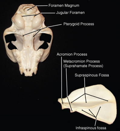
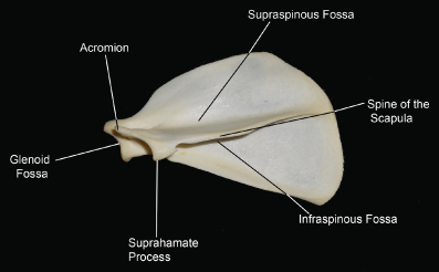
Conventionally, the scapula is considered to be a part of the limb because it is connected via the shoulder joint to the leg. The scapula forms the shoulder (scapulohumeral) joint in association with the humerus. The scapula is considered a flat bone. It does, however, have some additional geometry. The lateral surface of the scapula has a long, thin projection called the spine of the scapula (see Figure 3.4). It should be palpable on examination of the living animal. The ventral-most part of the spine of the scapula is called the acromion, which is important as a muscle attachment. The shallow depressions dorsal and ventral to the spine of the scapula are the supraspinous and infraspinous fossae, respectively. The fossa on the distal end of the scapula, into which the head of the humerus fits, is called the glenoid fossa. It is noteworthy that the scapula in cats and dogs is not directly attached to the rest of the skeleton; the only thing that holds it in place is muscle. Among other things, it means that it is easier to amputate the thoracic than the pelvic limb (such as in cases of malignancy) as no bony connections need be severed.
The humerus is the proximal-most long bone of the thoracic limb (see Figure 3.5). The humerus is distinguished by a head, a greater tuberosity, and a neck at its proximal boundary. The head of the humerus is a common site to sample bone marrow in cats. The humerus has a structure called the deltoid tuberosity on its medial surface, which is the attachment for the deltoid muscle. The distal humerus has two condyles, a lateral and a medial. In dogs, there is a trochlear foramen between the condyles. In cats, the equivalent is the supracondylar foramen, which is on the medial surface of the bone and proximal to the condyles rather than between them.
The humerus meets up with the radius and ulna at a joint that is referred to as the radiohumeral joint, and is commonly called the elbow. The radius actually crosses over the cranial surface of the ulna from the medial to the lateral side, while the ulna is situated so that it points slightly medially. The proximal ulna has a number of specific features of importance (see Figure 3.6). The olecranon is the part that projects caudally (or caudodorsally, depending on the position of the limb) and is clearly palpable when examining the leg during an office visit. Going distally, there is a C-shaped structure called the trochlear notch. The proximal tip of it is called the coronoid process, and the distal tip is called the anconeal process. There is a congenital abnormality in some dogs known as ununited anconeal process, which is difficult to diagnose, but is associated with lameness. There is a styloid process on the medial surface of the distal ulna.
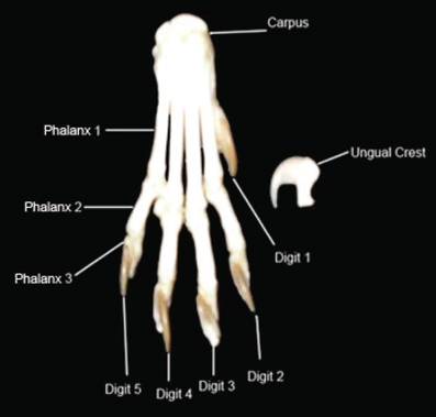
The joint distal to the elbow is the carpus, which is made of a series of short bones. Distal to those are bones that have been characterized as either flat or long bones, called the metacarpal bones. There is one for each digit. It is important to note that the metacarpals, and for that matter the digits, are numbered from the medial to the lateral side. In other words, the lateral-most digit in the dog and cat is number five. The dewclaw, which does not normally come in contact with the ground, is number one (see Figure 3.7).
The digits are a special case in that they are made of three bones in dogs and cats (usually only two in the dewclaw), at least one of which is irregular in shape, and one is short. Caudal to the metacarpophalangeal joints (the joints between the metacarpus and the proximal phalangeal bones) are small sesamoid bones, which help reduce tension on the tendons and ligaments of the paw.
The bones of the digits are called the proximal, middle, and distal phalangeal bones. They are often referred to as P1, P2, and P3, going distally. The distal part of P3 has a shallow fossa with a collar referred to as the ungual crest. It is here that the claw attaches to the digit (Figure 3.8).
Dogs may have dewclaws on all four limbs. Cats usually have dewclaws only on the thoracic limbs. Polydactyly is common in the dewclaw particularly, which is to say that there occasionally is more than one dewclaw on a given limb. It should be noted that pigs and some ruminants also have dewclaws.
The practice of removing the dewclaw was commonly done for hunting dogs. It was felt that the dewclaw could catch on the bushes and warn the prey animal that dogs and humans were in the area. Hunting dogs also often underwent docking, the removal of the distal tail, and cropping, the shortening of the pinna. Cropping was also done in show dogs. These procedures are illegal in Europe.
The Pelvic Limb
Unlike the thoracic limb, the joint where the pelvic limb connects to the body does, in turn, connect to the rest of the skeleton. As a result, the pelvic limb is considered to begin at the proximal-most long bone, which is the femur (see Figure 3.9). The femur is the longest bone in the dog and cat skeleton. It is distinguished by a large head, which fits into the coxofemoral (hip) joint. It has a prominent feature called the greater trochanter, which is on the lateral surface. It should be readily palpable on physical exam of the living animal. The distal end of the femur has a lateral and a medial condyle. The joint that is between the femur and the next most distal bone is the stifle joint; although the joint is properly called the femorotibial joint, the term stifle is universally used. It is incorrect to refer to this joint as the knee, as the knee in equines is actually the common name for the carpus, and confusion could easily result.
The bone distal to the stifle is the tibia (see Figure 3.10). The tibia has a plateau-like proximal surface; the small peak in its center is referred to as the intercondylar eminence. On its cranial surface, it has a projection called the tibial tuberosity. These landmarks are important in discussing surgery of the stifle joint, particularly involving cruciate ligament disorders.
One of the most striking characteristics of the stifle is the patella, which is actually contained within a tendon (the tendon of insertion of the quadriceps). It is the largest of all the sesamoid bones in mammals; its shape is considered to be like that of a sesame seed, which is where the entire category of these bones gets its name.
Running parallel to the tibia is the fibula, a thin bone that sits next to the tibia’s lateral surface. In equines and ruminants, this bone is actually fused to the tibia and is incomplete in its length. In dogs and cats, it ends along with the tibia at the next joint, the tarsus. This joint is made of a series of short and irregular bones. The tarsus has two irregular bones of note. One is the talus, which is rounded, and the other is the calcaneus, which is rectangular. The calcaneus is particularly important as the distal attachment for the gastrocnemius muscle, called the calcanean tendon; in humans, this is known as the Achilles tendon.
Distal to the tarsus are the metatarsal bones, distal to which are the digits. These bones are arrayed and numbered as they are in the thoracic limb. The exception, as mentioned, is that most cats do not have a dewclaw on the pelvic limbs.
Flat Bones
The Pelvis
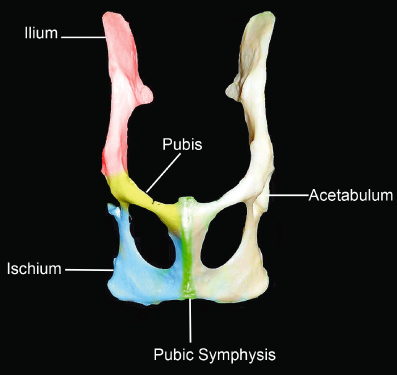
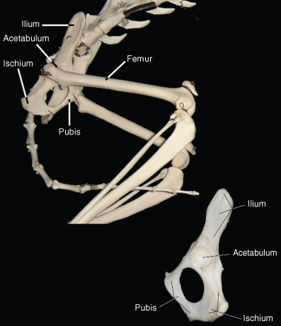
The pelvis is known as the os coxae (see Figure 3.11). Although it appears to be one large bone, it is actually composed of three flat bones that are fused during fetal development. The pelvis is considered part of the appendicular skeleton.
The cranial-most section is known as the ilium. The cranial part of the ilium sweeps dorsally and is called the wing of the ilium. A dorsocranial section of the wing is called the ileal crest and is important as an area from which we take bone marrow samples in dogs. (In cats, the bone is too thin, and the head of the humerus is used more often for this.)
The ischium is the section caudal and ventral to the ilium. It is a relatively short section but is important because it accommodates a fossa known as the acetabulum. This area is where the head of the femur sits, forming the coxofemoral joint. Fracture of the ischium, therefore, is often associated with hip joint problems. It should be noted that the acetabulum actually contains overlapping sections of the ilium, ischium, and pubis. Abnormalities of the acetabulum or of the head of the femur are part of hip dysplasia, which is a common orthopedic problem in large-breed dogs (see Figures 3.12 and 3.13).
The caudal part of the pelvis is the pubic bone. The pubis and the ischium together make up the borders of the obturator foramen, a large opening that is an important landmark.
The Skull
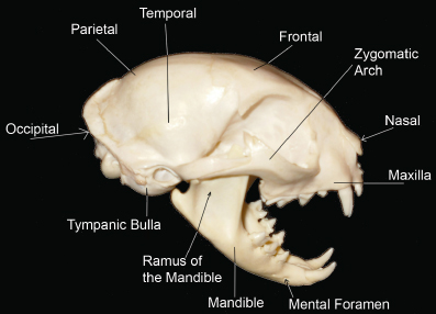
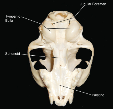
The skull is composed mostly of flat bones (see Figures 3.14–3.16). The cranium is the part of the skull that contains the brain. The jaw is composed of a single bone ventrally, called the mandible. The mandible has a dorsoventral projection at its caudal end that is called the ramus. It is the only part of the mandible that does not have teeth. The dorsal jaw is made of two bones that fuse together during development but is generally referred to by one name, the maxilla. Technically, the maxilla is the part that encompasses the lateral surface of the dorsal jaw. The rostral point of the upper (maxillary) jaw is composed of a small bone called the incisive bone, which holds the small teeth known as incisors.
The entire skull is made of a series of flat bones that eventually meet and become one structure. This happens during fetal development. Occasionally, when the dorsal plates of the skull have not completely come together at birth, there is a small spot on the dorsal cranium that is left with no bony covering. This is called a fontanel. It should close shortly after birth. It can allow significant damage to the brain by traumatic injury if it remains open.
The bone that is deep to the soft tissue that forms the roof of the mouth is the palatine bone. It too is composed of two bones that meet at the midline during fetal development. If these bones do not meet, the animal is born with a gap in the roof of the mouth, and milk or other ingested material can travel up into the nasal cavity and out the nose. This gap is referred to as a cleft palate and can usually be remediated surgically.


