© 2015 Mac Keith Press
6 Market Road, London, N7 9PW
Managing Director: Ann-Marie Halligan
Production Manager and Commissioning Editor: Udoka Ohuonu
Project Management: Lumina Datamatics
The views and opinions expressed herein are those of the authors and do not necessarily represent those of the publisher.
All rights reserved. No part of this publication may be reproduced, stored in a retrieval system, or transmitted in any form or by any means, electronic, mechanical, photocopying, recording or otherwise, without the prior written consent of the copyright holder or the publisher.
First published in this edition in 2015
British Library Cataloguing-in-Publication data
A catalogue record for this book is available from the British Library
Cover design: Karl Hunt
ISBN: 978-1-909962-53-8
Typeset by Lumina Datamatics, Chennai, India
Printed by Imprint Digital Limited, Rickmansworth, UK
Mac Keith Press is supported by Scope
CONTENTS
AUTHORS’ APPOINTMENTS
FOREWORD
PREFACE
1PLACENTAL PATHOLOGY IN UNDERSTANDING PERINATAL BRAIN INJURY
2ABNORMAL PLACENTAL PHENOTYPES
3ABERRANT PLACENTAL ENDOCRINOLOGY, FETAL GROWTH AND NEURODEVELOPMENT
4THE RHEOLOGY OF UTERO-PLACENTAL AND FETO-PLACENTAL BLOOD FLOW
5INFLAMMATION AND PLACENTATION
6INFECTIONS AND THE FETAL INFLAMMATORY RESPONSE
7CEREBRAL ISCHAEMIA AND WHITE MATTER INJURY
8IN UTERO IMAGING OF THE HUMAN PLACENTA
9CEREBRAL FUNCTION AND FETAL GROWTH RESTRICTION
10PLACENTAL PROGRAMMING AND MENTAL ILLNESS: FETAL GROWTH AND SCHIZOPHRENIA
11CEREBRO-THERAPEUTICS
12SUMMING-UP AND UNSOLVED PROBLEMS
INDEX
COLOR PLATE SECTION
AUTHORS’ APPOINTMENTS
| Kathryn Abel | Professor of Psychological Medicine, University of Manchester; Honorary Consultant Psychiatrist, Manchester Mental Health & Social Care Trust, Manchester, UK |
| Matthew Allin | Honorary Senior Lecturer, Institute of Psychiatry, King’s College London, UK |
| Martin Bax | Emeritus Reader in Child Health, Imperial College London, UK |
| Valentina Brusati | Specialist Surgeon in Obstetrics and Gynaecology, Department of Women, Mothers and Neonates, Buzzi Children’s Hospital; Institute of Clinical Specialisation, Milan, Italy |
| Irene Cetin | Head of Department of Mother and Child, Hospital Luigi Sacco; Director Giorgio Pardi Centre for Fetal Research Department of Biomedical and Clinical Sciences, University of Milan, Italy |
| Jayne Charnock | Post-Doctoral Research Associate, Maternal and Fetal Health Research Centre, Institute of Human Development, University of Manchester, UK |
| Ian Crocker | Senior Scientist/Lecturer, Maternal and Fetal Health Research Centre, Institute of Human Development, University of Manchester, UK |
| Michelle Desforges | Post-Doctoral Research Associate, Maternal and Fetal Health Research Centre, Institute of Human Development, University of Manchester; Manchester Academic Health Sciences Centre, UK |
| Emma Ingram | Clinical Research Fellow, Maternal and Fetal Health Research Centre, Institute of Human Development, University of Manchester, UK |
| Catherine James | Clinical Research Fellow, Maternal and Fetal Medicine, Institute for Women’s Health, University College London, UK |
| Ed Johnstone | Clinical Senior Lecturer, Institute of Human Development, University of Manchester, UK |
| Gil Mor | Professor of Obstetrics, Gynaecology and Reproductive Sciences, School of Medicine, Yale University, Connecticut, USA |
| Karin Nelson | Scientist Emeritus, National Institute of Neurological Disorders and Stroke, National Institutes of Health, Maryland, USA |
| Donald Peebles | Chair and Head of the Research Department of Maternal Fetal Medicine, University College London; Honorary Consultant in Maternal Fetal Medicine, Institute for Women’s Health, University College London Hospitals, UK |
| Karen Racicot | Assistant Professor, Department of Obstetrics, Gynecology and Reproductive Biology, College of Human Medicine, Michigan State University, USA |
| Raymond W Redline | Professor of Pathology, Case Western Reserve University; University Hospitals Case Medical Center, Ohio, USA |
| Colin Sibley | Professor of Child Health and Physiology, Maternal and Fetal Health Research Centre, Institute of Human Development, University of Manchester, UK |
| Philip Steer | Emeritus Professor of Obstetrics and Gynaecology, Imperial College London, UK |
| Suresh Victor | Senior Attending Physician, Neonatology Center of Excellence, Sidra Medical and Research Center, Doha, Qatar |
| Michael Weindling | Emeritus Professor of Perinatal Medicine, Department of Women’s and Children’s Health, University of Liverpool, UK |
| Melissa Westwood | Professor of Endocrinology, Maternal and Fetal Health Research Centre, Institute of Human Development, University of Manchester, UK |
FOREWORD
The development of the human brain in fetal life is of critical importance for lifelong function. Many neurodevelopmental deficits originate in the perinatal period and there is increasing awareness of the need to look into early life, including prenatal life, to understand the origins of cognitive development, healthy ageing and risk of neurodegenerative and psychiatric diseases.
The first edition of this book, edited by Philip Baker and Colin Sibley and published in 2006, drew attention to the role of utero-placental function as an important mediator of healthy fetal brain development. Since then there have been significant advances in this topic from a range of disciplines, including epidemiology, histopathology, endocrinology, imaging science, immunology, neuroprotection studies and rigorous assessment of clinical outcomes. Consequently, publication of a second edition that encompasses the new breadth of understanding is important and timely. This book succeeds unequivocally in addressing this need.
The book is structured in a logical way from pathology to clinical outcome. Throughout, information from the basic sciences is placed within the clinical context and there is excellent use of illustrative figures and images. These aspects of design and structure ensure that the volume is readily accessible to a broad readership.
All of the chapters are valuable. Redline’s chapter on placental pathology and perinatal brain injury sets the tone superbly and Sibley and Desforges continue to provide a detailed unifying theory of aberrant placentation and development, offering explanations for inadequate placental function and fetal compromise. The chapters by Charnock and Westwood, Crocker, and Racicot and Mor consider the roles of endocrine systems, placental haemodynamics and inflammation respectively, in placentation and function. There follows an excellent contribution from Peebles and James on infection and the fetal inflammatory response. Victor and Weindling discuss the synergy between inflammation and hypoxia-ischaemia as mediators of white matter injury and Cetin and Brusati discuss the problem of disturbed cerebral energetics in fetal growth restriction. There follow chapters on state-of-the-art methods for assessing placental structure, function and perfusion as well as fetal well-being using magnetic resonance imaging and ultrasound (Ingram and Johnstone), and magnetic resonance spectroscopy (Cetin and Brusati). Abel and Allin present the compelling case for the importance of prenatal factors on the causal pathway to psychiatric disease, while Steer provides a thorough overview of current strategies for fetal neuroprotection. Dr Nelson’s summary and discussion of knowledge gaps about the role of the placenta in fetal brain development provide an elegant and thought-provoking final chapter to this volume.
The editors should be congratulated for bringing together a panel of expert authors from diverse fields and for editing a volume that is comprehensive and authoritative. It should prove to be a valuable resource for basic scientists, obstetricians, neonatologists and pathologists and all those engaged in understanding the complex interactions between placental function and fetal brain development, as a basis for improving health outcomes across the life-course.
Dr James P Boardman
MRC Centre for Reproductive Health
University of Edinburgh
UK
PREFACE
The original concept and first edition of this book on placenta and neurodisability was developed from a meeting organised by Martin Bax and Mac Keith Meetings, held in the UK almost a decade ago with the support of the Castang Foundation. Over the intervening years, the links between this unique organ and the human brain, in utero as well as beyond, have expanded, aided not least by the continued enthusiasm and evangelism of Dr Bax and like-minded individuals. With a second meeting in 2010, it became obvious that knowledge, understanding and certainty in this topic had progressed, alongside clinical and technical advances and that a reworking of the original volume was necessary and somewhat overdue. Many participants at this meeting have contributed to this new edition and thankfully have formalised their links to expedite clinical and research progress across international borders.
As highlighted, neurodisability relating to perinatal brain injury is recognised in 2 to 3 of every 1000 live births. However, the numbers associated with more subtle, late-onset neurophysical disorders, perhaps exacerbated by environmental factors, may be more profound. As is evident throughout the text, improvements in obstetric care and neonatal surveillance have served to increase infant survival, particularly for those of very low birthweight and defined contributory lesions. Nevertheless, iatrogenic stresses remain a concern, with overriding importance given to developmental immaturity, underlying fetal growth restriction, cardiopulmonary instability and infections, placental or otherwise. In many cases, placental pathology provides a circumscribed role, identifying inflammatory responses and functional deficits. Nevertheless, for cases of neurodisability at term or in the near-term infant, it is becoming increasingly clear, as indicated in this book, that the placenta has a major role in the pathophysiology which underscores, or at the very least contributes, to central nervous system injury and dysplasia.
To this end, this book covers all necessary components and discussion points regarding these placental contributions, from inflammation (infectious and immunological) through aberrations in placental and fetal growth, the role of genetic and environmental factors in neurological development, along with current clinical approaches to monitoring and restricting aberrations. Nevertheless, as emphasised by Karin Nelson in the final chapter, more work lies ahead. Along with clarification of the fetal-placental insults which beget developmental issues, there is a specific need for follow-up studies in humans to delineate disorders, strenghthen associations with placental pathophysiology and define the biological foundations of congenital neurodisability. Only when these are determined will progress in preventing and offsetting this string of pathological events be forthcoming, with a possible leap forward in fetal and perinatal medicine. Nonetheless, as a marker of current knowledge in all aspects of this complex story, this book stands alone in its breadth and accessibility, to be read by experts and inquisitive minds alike.
Ian Crocker
Maternal and Fetal Health Research Centre
University of Manchester, UK
April 2015
1
PLACENTAL PATHOLOGY IN UNDERSTANDING PERINATAL BRAIN INJURY
Raymond W Redline
Introduction
Static disorders of motor function, such as cerebral palsy (CP), develop within the first two years of life in approximately 2 – 3/1000 children (Himmelmann et al. 2010). Depending on the regional prevalence of premature birth (especially at less than 28 weeks gestation), the proportion of cases related to extreme immaturity may vary from between 25 and 50%. In almost all cohorts the proportion of otherwise unremarkable term infants (> 37 wks) is relatively constant at around 40% – 50%. The remaining cases are a heterogeneous group, including near term infants, markedly growth restricted infants of all gestational ages, infants with undiagnosed genetic or chromosomal disorders and infants with neurotoxic exposures to infectious agents or other toxins in either the prenatal or postnatal period.
Recognized clinical risk factors for CP and related disorders in the extremely preterm infant include earlier gestational age, superimposed fetal growth restriction, a low Apgar score at birth, postnatal hypoxia, overly aggressive ventilation with hyperoxia–hypocarbia, patent ductus arteriosus, postnatal corticosteroid therapy and postnatal inflammatory disorders including necrotizing enterocolitis and late onset sepsis (Babcock et al. 2009). The degree of risk associated with ascending bacterial infections (chorioamnionitis), as discussed below, remains controversial and is considered in Chapter 6. Recognized risk factors for central nervous system (CNS) injury in term infants include a positive family history of neurodevelopmental disorders, fetal growth restriction, abnormal neurological examination in the first days of life (neonatal encephalopathy) and hypothyroidism (Redline 2008a). The degree of risk associated with so-called birth asphyxia (recently defined by several international consensus conferences) is also controversial and will be discussed later in the book. Novel therapies decreasing the susceptibility of neurons to injury, including magnesium sulfate in preterm infants and head cooling in term infants, raise the possibility of attenuating CNS injury associated with acute insults, but may be less efficacious in cases with coexisting placental pathology.
The existing literature on placental pathology and CNS injury suffers from several problems. First, adverse outcomes are rare and often diagnosed years after birth. Other limitations include bias of ascertainment, lack of an appropriate control group, inadequate characterization of placental lesions, use of surrogate short-term outcomes, unmeasurable differences in genetic susceptibility to injury and failure to fully account for the effects of gestational age and post birth complications. Nevertheless, contrary to the conclusions of a recent commentary (Nelson and Blair 2011), strong working hypotheses have emerged from the large number of published reports regarding which types of placental lesions are most likely to increase the risk of CNS injury. The focus of this chapter will be to place these pathologic findings in perspective in terms of their ability to cause or alter the threshold for the various manifestations of perinatal brain injury including abnormal neuroimaging, neonatal encephalopathy, ischemic stroke, seizure disorders and later disabilities such as CP and developmental delay in live born infants and neuropathological changes in stillbirths.
Potential placental mechanisms for injury
The placenta is essential for fetal life; constituting its only source for oxygen, water, nutrients and elimination of waste products. So in a sense it is not surprising that placental dysfunction can lead to disordered growth and development or injury to specific organs such as the brain and spinal cord. However, it has been challenging to isolate the particular patterns of placental injury that are most likely to cause damage. Before considering specific placental lesions, the potential pathways by which placental dysfunction could affect fetal brain function will be briefly reviewed.
PAUCITY OF PROTECION
In this scenario the fetus is deprived of specific crucial elements such as essential amino acids, maternal or placental hormones, or minerals and vitamins that are necessary for CNS development or protection from injury (Dammann and Leviton 1999). One simple way this can occur is preterm birth, that is, severing the fetus from its maternal supply line. Other potential mechanisms include genetic or epigenetic abnormalities in placental transporters or growth factor expression and maternal deficiencies in dietary intake or metabolic state. While these pathways may be extremely important, as evidenced by the importance of a positive family history and the known effects of maternal hypothyroidism and phenylketonuria on later childhood CNS function, they have no known structural correlate and cannot be detected by pathological examination.
DYSFUNCTION OF CORE PATHWAYS
The supply line between mother and fetus depends on adequate maternal circulating volume and blood pressure; an intact and appropriately remodeled utero-placental vasculature; free circulation through and drainage of the placental intervillous spaces; a short diffusion distance between maternal blood and fetal capillaries; and an intact, non-obstructed feto-placental circulation (see Fig. 1.1). Pathophysiological processes directly affecting these core pathways fall into two categories. The first category includes acute sentinel events such as maternal shock, uterine rupture, abruptio placenta, umbilical cord occlusion and feto-placental hemorrhages. These often result in either fetal death or recovery without CNS sequelae, but in some cases lead to global asphyxia and later CP with major developmental disabilities (Myers 1975). The second category includes chronic sub-lethal processes leading to gradual loss of function and decreased placental reserve to withstand later injury (i.e. situations where fetal demand exceeds placental supply). Processes associated with decreased placental reserve include maternal malperfusion (MMP), secondary to inadequate maternal vascular remodeling, inadequate placental mass (placental weight less than 10th centile for gestational age) and processes affecting the diffusing capacity across the interhemal membrane (placental maturation defect, perivillous fibrin(oid) deposition and villitis of unknown etiology [VUE]), that is unexplained villus infiltration by maternal T lymphocytes.
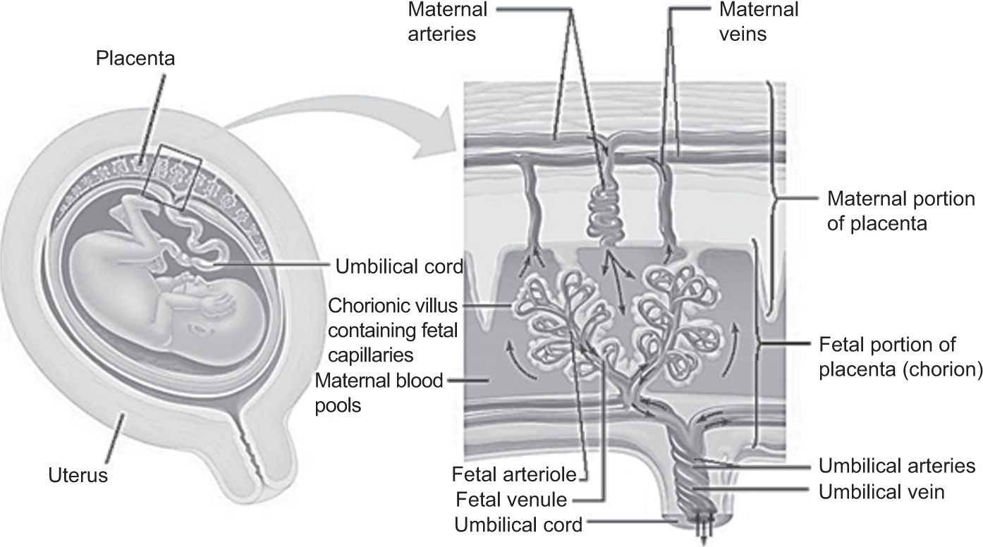
Fig. 1.1. Topological depiction of the human placenta, showing main structural elements and maternal and fetal blood flow. Reproduced by permission Pearson Education © 2013.
DISTURBANCES INVOLVING MULTIPLE DISTINCT PHYSIOLOIC COMPARTMENTS
Numerous studies have shown that CNS injury is strongly associated with multiple placental lesions (so-called ‘mixed pathology’). These lesions may act synergistically to disrupt a single core pathway or in parallel by blocking both primary and compensatory pathways. They can act either directly by affecting gas exchange or indirectly via the elaboration of harmful circulating mediators including cytokines, pathogen or damage associated molecular patterns (PAMP/DAMP), complement fragments, microparticles, activated coagulation components, vasoactive molecules and reactive oxygen species. It is important to distinguish the finding of multiple independent lesions from multiple abnormalities that all characterize a single pattern of injury (so-called constellation disorders).
PRECONDITIONING INVOLVING PLACENTAL LESIONS OCCURING AT DIFFEREENT TIMES
The general hypothesis of preconditioning is that changes of state induced by prior events can either positively or negatively modulate the deleterious effects of a subsequent insult (Hagberg et al. 2004). In the placenta these sequential insults generally involve repeated exposure to hypoxia, circulating inflammatory mediators, or some combination of the two. However, they could also potentially include placental conditions that alter the fetal endocrine or metabolic state. The concept of preconditioning helps to explain the inconsistent association of any single placental lesion with CNS injury. Since preconditioning can also be positive, this may explain why in some studies processes such as chorioamnionitis and mild MMP have actually been found to be neuroprotective.
Placental pathology in CNS injury
Key characteristics of a good pathological study include the use of clearly defined and generally accepted findings that characterize interruption of a specific physiological pathway, incorporation of scaling that distinguishes the intensity (grade), duration (stage), extent and character of these findings and comparison with an appropriate control group. Ideally specific criteria for each finding should be fully described as reproducibility of these findings is an important consideration for clinical application and studies involving multiple different scorers. However this is not a central issue for studies undertaken in a single center or where scoring is done at a central reading center. Unfortunately, few studies have met all of these benchmarks. This, combined, with the logistic issues and clinical confounders mentioned above, contributes to continuing skepticism regarding the validity of placental pathology in helping to explain adverse neurological outcomes.
PATTERNS ASSOCIATED WITH CNS INJURY AT ALL GESTATIONAL AGES
Several types of placental injury have been consistently associated with different types of CNS injury in diverse clinical scenarios at all gestational ages. The identification of more than one placental lesion has been the strongest and most consistent predictor between studies and is the finding least influenced by other risk factors and confounders (Redline and O’Riordan 2000, Viscardi and Sun 2001, Chang et al. 2011). Also, the degree of risk has been shown to increase significantly with each additional placental lesion identified. The most commonly observed combinations are a vascular lesion (either maternal or fetal) with an inflammatory process (either acute or chronic); however combinations of a maternal and fetal vascular lesion or acute and chronic inflammation are also seen.
A second set of findings in diverse types of CNS injury are placental changes indicative of severe MMP, such as villous infarction (Fig. 1.2a). These have been associated with neuronal necrosis in stillborn fetuses, CP and developmental delay in preterm infants and CP in association with neonatal encephalopathy in term infants (Burke and Tannenberg 1995, Redline et al. 2007). A much less common form of maternal perfusion abnormality known as maternal floor infarction has also been associated with both motor disorders and developmental delay at all gestational ages (Adams-Chapman et al. 2002).
Finally, pathological findings suggestive of chronic partial or intermittent umbilical cord obstruction, have also been implicated in several studies of CNS injury (Grafe 1994, Redline 2008b). The biological plausibility for this pattern is supported by neurological follow-up in infants with sonographic evidence of persistent cord entanglements and animal studies documenting the effects of repetitive episodes of fetal hypoxia and partial prolonged asphyxia (Myers 1975, Clapp et al. 2003). Gross pathological lesions affecting the umbilical cord, such as membranous insertion, excessive length, increased coiling and tight knots can all potentially restrict feto-placental blood flow. When combined with histological indicators, such as scattered small foci of avascular villi, intimal fibrin cushions in stem villous vessels, perivascular edema around stem villous arteries, distal villous immaturity and chorionic/stem villous venous dilation at the cord insertion site, they can cause a physiologically significant reduction in umbilical blood flow, increased placental venous pressure and fetal vascular stasis.
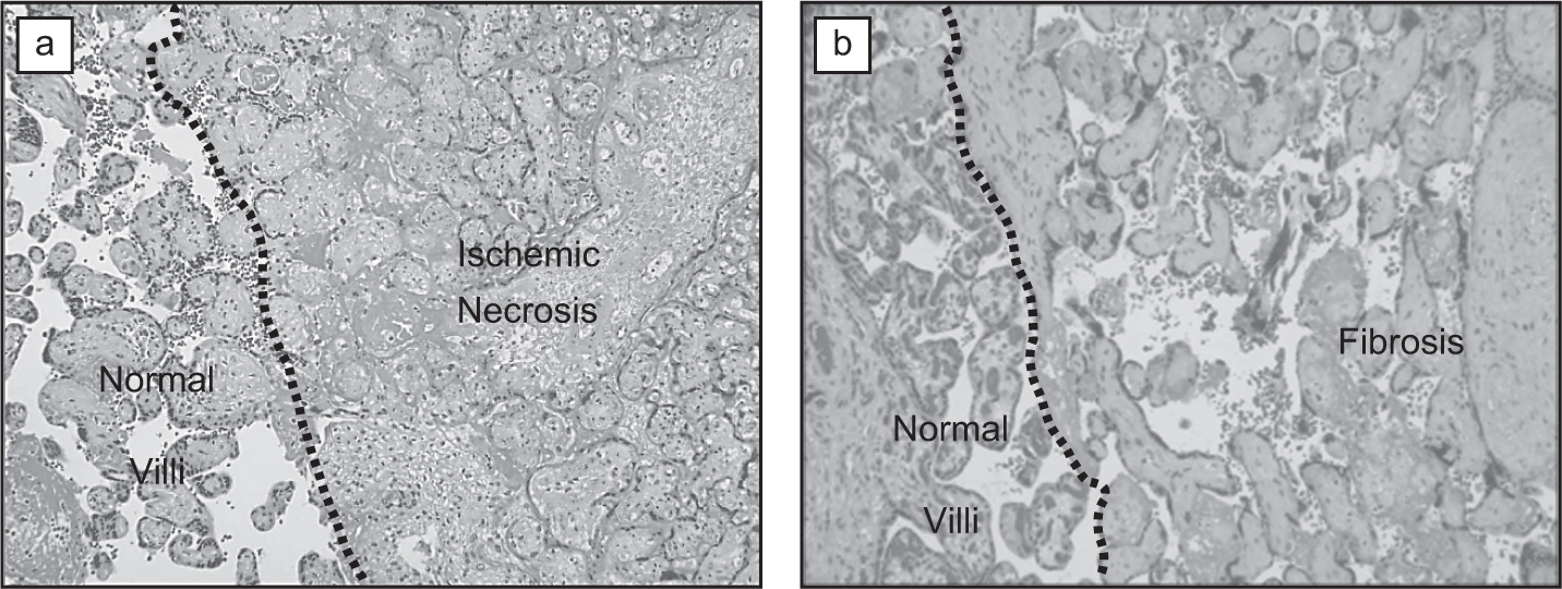
Fig. 1.2. Placental vascular lesions with high prevalence in neonatal brain injury. (a) Maternal villous infarct: A portion of the distal villous tree shows ischemic necrosis of villous trophoblast and obliteration of the intervillous space, secondary to obstruction of a maternal spiral artery. Villous stromal and vascular architecture is preserved. Normal villi are seen on the left (H&E stain, 10x magnification). (b) Fetal thrombotic vasculopathy: A portion of the distal villous tree shows loss of fetal vessels and stromal fibrosis secondary to obstruction of a large fetal placental vessel. Villous trophoblast and intervillous space are preserved. Normal villi seen on the left side (H&E stain, 10x magnification). A colour version of this figure can be seen in the plate section at the end of the book.
PATTERNS ASSOCIATED WITH CNS INJURY IN PRETERM INFANTS
The majority of studies addressing CNS injury in preterm infants have focused on histologic chorioamnionitis (HCA). Although meta-analysis has found HCA to be a significant risk factor for CP, the exact nature of the relationship remains unclear (Shatrov et al. 2010). Several specific problems confound this association: (1) the prevalence of HCA increases dramatically with decreasing gestational age, the strongest predictor of CNS injury in this population; (2) HCA is highly prevalent in this population (over 50%) and often seen with precipitous uncontrolled vaginal deliveries, while other less frequent causes of preterm birth (twins, pre-eclampsia) are often more closely monitored and more likely to undergo elective operative deliveries; (3) HCA-negative comparison groups are enriched for placentas with mild MMP, which may exert a protective effect against CNS injury (see below); (4) HCA predisposes to post-birth neonatal complications that may themselves be the cause of injury; and (5) HCA is a complex mixture of different grades and stages of maternal and fetal inflammation that have not been adequately separated in many studies.
In placentas with HCA the strongest and most consistent risk factor for CNS injury is acute fetal vasculitis affecting either umbilical (funisitis) or chorionic plate (chorionic vasculitis) vessels. Fetal vasculitis is not a true vasculitis, but rather transmigration of fetally-derived neutrophils toward bacteria in the amniotic fluid. When found in umbilical arteries, compared with the umbilical vein alone, fetal vasculitis has been correlated with elevated levels of fetal cytokines (Rogers et al. 2002). Specific outcomes in preterm infants that have been associated with HCA with or without FV include intraventricular hemorrhage, white matter lesions on early head ultrasound and developmental delay at various periods during childhood (Leviton et al. 1999, Mittendorf et al. 2003). Other studies have shown that increased intensity of FV, recent non-occlusive fetal thrombi and maternal perfusion abnormalities all increase the risk of injury with HCA (Redline et al 1998, Kaukola et al. 2006). However, a few studies have found that HCA is associated with a decreased risk of death and no change in the risk of neurological impairment, illustrating the continuing controversy regarding the exact nature of this relationship (Dexter et al. 2000, Andrews et al. 2008).
Additional placental lesions that have been associated with more than one pattern of CNS injury in preterm infants include diffuse villous edema, severe MMP and two decidual lesions, recent hemorrhage and fibrin and lymphoplasmacytic inflammation (Kumazaki et al. 2002, Mehta et al. 2006, Redline et al. 2007, Maleki et al. 2009, Leviton et al. 2010). Interestingly, mild MMP was associated with a reduced risk of adverse outcomes in one study (Redline et al. 1998).
PATTERNS ASSOCIATED WITH CNS INJURY IN TERM INFANTS
Methodological issues such as the inability to identify infants at risk and a lack of appropriate controls, have made the study of placental pathology and CNS injury in term infants particularly difficult. Observations in specific cohorts such as cases in litigation, infants with neonatal encephalopathy, tertiary care referrals for head cooling and stillbirths with detailed neuropathologic examination provide some insight. Fetal thrombotic vasculopathy (FTV, Fig. 1.2b) and other pathological processes damaging large feto-placental vessels in the umbilical cord and/or chorionic plate (VUE with obliterative fetal vasculopathy, meconium-associated fetal vascular necrosis and HCA with intense fetal vasculitis) have emerged as highly prevalent and strongly significant predictors of CNS injury. In the largest studies, these have been specifically associated with neuronal damage, neonatal encephalopathy and CP (McDonald et al. 2004, Redline 2005, Chang et al. 2011). Although often seen in cases of intrapartum distress, their effects appear to be independent of indicators of birth asphyxia such as low cord pH and 5 minute Apgar score. In term infants referred for head cooling they have been found by some, but not all, investigators to predict a decreased responsiveness to therapy compared with infants showing evidence of birth asphyxia alone (Wintermark et al. 2010, Ernst et al. 2012).
Other pathologic lesions associated with CNS injury in term placentas include heterochronic placental lesions (one present for weeks, the other present for 6 – 12 hours or more), scattered foci of avascular villi suggestive of umbilical cord obstruction and placental infarcts (Redline and O’Riordan 2000, Redline 2008, Nelson and Blair 2011). In smaller studies, placental maturation defect and FTV were found to be more prevalent in children with perinatal stroke, whilst placental maturation defect and VUE were more prevalent in infants with seizures (Scher et al. 1998, Elbers et al. 2011).
Pathological approach
Finally, leaving theoretical and epidemiological considerations aside, I will briefly describe how I, as a pathologist and medico-legal consultant, approach the relationship between specific placental lesions and CNS injury in individual cases. I generally subdivide cases into six categories. These categories are hierarchical in the sense that earlier categories take precedence over later ones. However, the finding of lesions in multiple categories is very common and undoubtedly increases the risk of significant CNS damage.
1: LESIONS SUFFICIENT ALONE TO EXPLAIN CNS INJURY
These are the so-called ‘sentinel events’ such as abruptio placenta, uterine rupture, fetal hemorrhage, or complete umbilical cord obstruction. Since these lesions cause critical hypoxia they are almost by definition acute, occurring within a period of minutes to hours before birth. Placental findings consistent with sentinel events are generally identified at gross examination and include large retroplacental hematomas, torn fetal vessels, or tight umbilical cord knots. Microscopic examination is generally supplementary to the clinical history and gross examination in these situations. Placental evaluation can sometimes change the clinical diagnosis of a sentinel event by failing to substantiate the clinical impression and/or by identifying alternative explanations.
2: LESIONS SUFFICIENT ALONE TO EXPLAIN CNS INJURY, BUT USUALLY SEEN IN COMBINATION WITH INTRAPARTUM DISTRESS
The main diagnoses in this category are the thrombotic and inflammatory lesions that affect large fetal placental blood vessels including FTV, VUE with obliterative fetal vasculopathy and prolonged meconium exposure with vascular necrosis. Another inflammatory lesion, chorioamnionitis with an intense fetal vasculitis, has been reported as a risk factor in some studies, particularly when combined with recent non-occlusive chorionic vessel thrombi in preterm infants. These types of lesions not only affect placental function, but can also compromise fetal circulatory physiology and directly cause fetal organ damage via the effects of circulating mediators such as cytokines, microparticles and activated coagulation components.
3: LESIONS SUFFICIENT TO CAUSE INJURY, BUT OF LOWER SPECIFICITY
These lesions are also highly prevalent in cases of CNS injury, but overlap with findings observed in placentas submitted for other reasons. Three lesions in this category are chronic partial/intermittent umbilical cord obstruction, marked MMP and diffuse villous edema. The first two are so-called ‘constellation disorders’ that depend on the identification of multiple related findings. In these processes it can be difficult to decide which combinations of findings are most specific for significant placental dysfunction. Further studies are needed to dissect patterns and severities that allow more accurate prediction. One finding that can assist in assessing the degree of associated hypoxia is an elevation in the number of immature red blood cell precursors in the fetal placental circulation (see below). The third lesion in this category, diffuse villous edema, is only seen in the placentas of extremely low gestational age preterm infants. There is some ambiguity related to this lesion since it is somewhat subjective and an exaggeration of a normal developmental pattern seen in late second trimester placentas. Nevertheless, it has been a strong predictor across studies and is independent of gestational age by multivariate analysis.
4: LESIONS USUALLY NOT SUFFICIENT ALONE TO CAUSE INJURY, BUT SYNERGISTIC WITH INTRAPARTUM STRESS
Lesions in this category decrease the placental reserve by compromising the efficiency of placental function. While not directly causal, they can decrease the threshold for comparatively minor episodes of perinatal stress to compromise CNS function. Most cases of MMP, distal villous immaturity/placental maturation defect, VUE without obliterative vasculopathy, chronic abruption and perivillous fibrinoid deposition without maternal floor infarction fall within this category.
5: NON-CAUSAL ADAPTIVE LESIONS INDICATIVE OF ANTENATAL STRESS
There are two lesions in this category: villous chorangiosis and increased circulating fetal nucleated red blood cells (NRBC). Chorangiosis reflects increased villous angiogenesis developing over weeks in response to decreased maternal oxygen tension with normal maternal perfusion. Recognized associations include maternal anemia, smoking and pregnancy at high altitudes (Ogino and Redline 2000). Significantly increased circulating NRBC (more than one per high power field, corresponding to a neonatal count of greater than 2500/mm3) represent a physiologic response to decreased fetal oxygen tension with mobilization of immature red blood cell precursors from intramedullary and extramedullary sources (Redline 2008b). Significantly elevated NRBC counts are believed to develop after a period of at least 6–12 hours of significant tissue hypoxia.
6. NO IDENTIFIABLE PLACENTAL LESION
This final category is heterogeneous. Explanations to consider when placentas show no significant lesions include unrecognized clinical sentinel events, birth trauma, inadequate placental examination and intrinsic CNS susceptibility due to genetic or epigenetic abnormalities. The latter explanation is most plausible when a positive family history of neurodevelopmental disorders exists.
Conclusions
Extrinsic factors aside, a unifying hypothesis to account for the relationship between the placenta and CNS injury is that, between 23 and 39 weeks’ gestation there exists a critical period for brain development and resilience to injury that requires a fully functioning placenta. Birth before this stage (paucity of protection) and placental dysfunction during this stage (decreased reserve and preconditioning) can decrease the threshold for problems in the perinatal period. These placental processes also interact with inherent variations in the set point for CNS injury, a variable that is likely to be influenced by poorly understood genetic and epigenetic factors. Together with better understanding of relevant physiology and genetics and better epidemiological studies, the development of antenatal screening tests to identify fetuses and placentas at risk is a major goal for the prevention, early diagnosis and prompt treatment of perinatal brain damage.
REFERENCES
Adams-Chapman I, Vaucher YE, Bejar RF, Benirschke K, Baergen RN, Moore TR (2002) Maternal floor infarction of the placenta: Association with central nervous system injury and adverse neurodevelopmental outcome. J Perinatol 22(3): 236–241. doi: http://dx.doi.org/10.1038/sj.jp.7210685
Andrews WW, Cliver SP, Biasini F et al. (2008) Early preterm birth: Association between in utero exposure to acute inflammation and severe neurodevelopmental disability at 6 years of age. Am J Obstet Gynecol 198(4): 466.e1–466.e11. doi: http://dx.doi.org/10.1016/j.ajog.2007.12.031
Babcock MA, Kostova FV, Ferriero DM et al. (2009) Injury to the preterm brain and cerebral palsy: Clinical aspects, molecular mechanisms, unanswered questions, and future research directions. J Child Neurol 24(9): 1064–1084. doi: http://dx.doi.org/10.1177/0883073809338957
Blair E, de Groot J, Nelson KB (2011) Placental infarction identified by macroscopic examination and risk of cerebral palsy in infants at 35 weeks of gestational age and over. Am J Obstet Gynecol 205(2): 124.e1–124.e7. doi: http://dx.doi.org/10.1016/j.ajog.2011.05.022
Burke CJ, Tannenberg AE (1995) Prenatal brain damage and placental infarction: An autopsy study. Dev Med Child Neurol 37: 555–562. doi: http://dx.doi.org/10.1111/j.1469-8749.1995.tb12042.x
Chang KT, Keating S, Costa S, Machin G, Kingdom J, Shannon P (2011) Third-trimester stillbirths: Correlative neuropathology and placental pathology. Pediatr Dev Pathol 14(5): 345–352. doi: http://dx.doi.org/10.2350/10-07-0882-oa.1
Clapp JF III, Stepanchak W, Hashimoto K, Ehrenberg H, Lopez B (2003) The natural history of antenatal nuchal cords. Am J Obstet Gynecol 189(2): 488–493. doi: http://dx.doi.org/10.1067/s0002-9378(03)00371-5
Dammann O, Leviton A (1999) Brain damage in preterm newborns: Might enhancement of developmentally regulated endogenous protection open a door for prevention? Pediatrics 104(3 Pt 1): 541–550.
Dexter SC, Pinar H, Malee MP, Hogan J, Carpenter MW, Vohr BR (2000) Outcome of very low birth weight infants with histopathologic chorioamnionitis. Obstet Gynecol 96(2): 172–177. doi: http://dx.doi.org/10.1016/s0029-7844(00)00886-3
Elbers J, Viero S, MacGregor D, deVeber G, Moore AM (2011) Placental pathology in neonatal stroke. Pediatrics 127(3): e722–e729. doi: http://dx.doi.org/10.1542/peds.2010-1490
Ernst LM, de Regnier RO, Boswell L, Huang MH, Khan JY (2012) Clinicopathologic factors as predictors of outcome in term infants with hypoxic ischemic encephalopathy undergoing therapeutic hypothermia. Pediatr Dev Pathol 14: 510–511.
Grafe MR (1994) The correlation of prenatal brain damage with placental pathology. Neuropathol Exp Neurol 53: 407–415. doi: http://dx.doi.org/10.1097/00005072-199407000-00013
Hagberg H, Dammann O, Mallard C, Leviton A (2004) Preconditioning and the developing brain. Semin Perinatol 28(6): 389–395. doi: http://dx.doi.org/10.1053/j.semperi.2004.10.006
Himmelmann K, Hagberg G, Uvebrant P (2010) The changing panorama of cerebral palsy in Sweden. X. Prevalence and origin in the birth-year period 1999–2002. Acta Paediatr 99(9): 1337–1343. doi: http://dx.doi.org/10.1111/j.1651-2227.2010.01819.x
Kaukola T, Herva R, Perhomaa M et al. (2006). Population cohort associating chorioamnionitis, cord inflammatory cytokines and neurologic outcome in very preterm, extremely low birth weight infants. Pediatr Res 59(3): 478–483. doi: http://dx.doi.org/10.1203/01.pdr.0000182596.66175.ee
Kumazaki K, Nakayama M, Sumida Y et al. (2002). Placental features in preterm infants with periventricular leukomalacia. Pediatrics 109(4): 650–655. doi: http://dx.doi.org/10.1542/peds.109.4.650
Leviton A, Allred EN, Kuban KCK et al. (2010) Microbiologic and histologic characteristics of the extremely preterm infant's placenta predict white matter damage and later cerebral palsy. The ELGAN study. Pediatr Res 67(1): 95–101. doi: http://dx.doi.org/10.1203/pdr.0b013e3181bf5fab
Leviton A, Paneth N, Lynne Reuss M et al. (1999) Maternal infection, fetal inflammatory response and brain damage in very low birth weight infants. Developmental Epidemiology Network Investigators. Pediatr Res 46(5): 566–575. doi: http://dx.doi.org/10.1203/00006450-199911000-00013
Maleki Z, Bailis AJ, Argani CH, Askin FB, Graham EM (2009). Periventricular leukomalacia and placental histopathologic abnormalities. Obstet Gynecol 114(5): 1115–1120. doi: http://dx.doi.org/10.1097/aog.0b013e3181bdcfc4
McDonald DG, Kelehan P, McMenamin JB et al. (2004). Placental fetal thrombotic vasculopathy is associated with neonatal encephalopathy. Hum Pathol 35(7): 875–880. doi: http://dx.doi.org/10.1016/j.humpath.2004.02.014
Mehta R, Nanjundaswamy S, Shen-Schwarz S, Petrova A (2006). Neonatal morbidity and placental pathology. Indian J Pediatr 73(1): 25–28. doi: http://dx.doi.org/10.1007/bf02758255
Mittendorf R, Montag AG, MacMillan W et al. (2003). Components of the systemic fetal inflammatory response syndrome as predictors of impaired neurologic outcomes in children. Am J Obstet Gynecol 188(6): 1438–1446. doi: http://dx.doi.org/10.1067/mob.2003.380
Myers RE (1975) Four patterns of perinatal brain damage and their conditions of occurrence in primates. Adv Neurol 10: 223–234.
Nelson KB, Blair E (2011) The placenta and neurologic and psychiatric outcomes in the child: Study design matters. Placenta 32(9): 623–625. doi: http://dx.doi.org/10.1016/j.placenta.2011.06.021
Ogino S, Redline RW (2000) Villous capillary lesions of the placenta: Distinctions between chorangioma, chorangiomatosis and chorangiosis. Hum Pathol 31: 945–954. doi: http://dx.doi.org/10.1053/hupa.2000.9036
Redline RW (2005) Severe fetal placental vascular lesions in term infants with neurologic impairment. Am J Obstet Gynecol 192: 452–457. doi: http://dx.doi.org/10.1016/j.ajog.2004.07.030
Redline RW (2008a) Cerebral palsy in term infants: A clinicopathologic analysis of 158 medicolegal case reviews. Pediatr Dev Pathol 11(6): 456–464. doi: http://dx.doi.org/10.2350/08-05-0468.1
Redline RW (2008b) Elevated circulating fetal nucleated red blood cells and placental pathology in term infants who develop cerebral palsy. Hum Pathol 39(9): 1378–1384. doi: http://dx.doi.org/10.1016/j.humpath.2008.01.017
Redline RW, Minich N, Gerry Taylor H, Hack M (2007). Placental lesions as predictors of cerebral palsy and abnormal neurocognitive function at school age in extremely low birth weight infants (<1 kg). Pediatr Dev Pathol 10(4): 282–292. doi: http://dx.doi.org/10.2350/06-12-0203.1
Redline RW, O’Riordan MA (2000) Placental lesions associated with cerebral palsy and neurologic impairment following term birth. Arch Pathol Lab Med 124(12): 1785–1791.
Redline RW, Wilson-Costello D, Borawski E, Fanaroff AA, Hack M (1998). Placental lesions associated with neurologic impairment and cerebral palsy in very low birth weight infants. Arch Pathol Lab Med 122: 1091–1098.
Rogers BB, Alexander JM, Head J, Mcintire D, Leveno KJ (2002) Umbilical vein interleukin-6 levels correlate with the severity of placental inflammation and gestational age. Hum Pathol 33(3): 335–340. doi: http://dx.doi.org/10.1053/hupa.2002.32214
Scher MS, Trucco GS, Beggarly ME, Steppe DA, Macpherson TA (1998) Neonates with electrically confirmed seizures and possible placental associations. Pediatr Neurol 19: 37–41. doi: http://dx.doi.org/10.1016/s0887-8994(98)00012-5
Shatrov JG, Birch SC, Lam LT, Quinlivan JA, McIntyre S, Mendz GL (2010). Chorioamnionitis and cerebral palsy: A meta-analysis. Obstet Gynecol 116(2 Pt 1): 387–392. doi: http://dx.doi.org/10.1097/aog.0b013e3181e90046
Viscardi RM, Sun CC (2001). Placental lesion multiplicity: Risk factor for IUGR and neonatal cranial ultrasound abnormalities. Early Hum Dev 62(1): 1–10. doi: http://dx.doi.org/10.1016/s0378-3782(01)00114-1
Wintermark P, Boyd T, Gregas MC, Labrecque M, Hansen A (2010). Placental pathology in asphyxiated newborns meeting the criteria for therapeutic hypothermia. Am J Obstet Gynecol 203(6): 579.e1–579.e9. doi: http://dx.doi.org/10.1016/j.ajog.2010.08.024