

This edition first published 2020
© 2020 John Wiley & Sons, Inc.
All rights reserved. No part of this publication may be reproduced, stored in a retrieval system, or transmitted, in any form or by any means, electronic, mechanical, photocopying, recording or otherwise, except as permitted by law. Advice on how to obtain permission to reuse material from this title is available at http://www.wiley.com/go/permissions.
The right of Brian G. Murphy, Cynthia M. Bell and Jason W. Soukup to be identified as the authors of this work has been asserted in accordance with law.
Registered Offices
John Wiley & Sons, Inc., 111 River Street, Hoboken, NJ 07030, USA
Editorial Office
111 River Street, Hoboken, NJ 07030, USA
For details of our global editorial offices, customer services, and more information about Wiley products visit us at www.wiley.com.
Wiley also publishes its books in a variety of electronic formats and by print‐on‐demand. Some content that appears in standard print versions of this book may not be available in other formats.
Limit of Liability/Disclaimer of Warranty
The contents of this work are intended to further general scientific research, understanding, and discussion only and are not intended and should not be relied upon as recommending or promoting scientific method, diagnosis, or treatment by clinician for any particular patient. In view of ongoing research, equipment modifications, changes in governmental regulations, and the constant flow of information relating to the use of medicines, equipment, and devices, the reader is urged to review and evaluate the information provided in the package insert or instructions for each medicine, equipment, or device for, among other things, any changes in the instructions or indication of usage and for added warnings and precautions. While the publisher and authors have used their best efforts in preparing this work, they make no representations or warranties with respect to the accuracy or completeness of the contents of this work and specifically disclaim all warranties, including without limitation any implied warranties of merchantability or fitness for a particular purpose. No warranty may be created or extended by sales representatives, written sales materials or promotional statements for this work. The fact that an organization, website, or product is referred to in this work as a citation and/or potential source of further information does not mean that the publisher and authors endorse the information or services the organization, website, or product may provide or recommendations it may make. This work is sold with the understanding that the publisher is not engaged in rendering professional services. The advice and strategies contained herein may not be suitable for your situation. You should consult with a specialist where appropriate. Further, readers should be aware that websites listed in this work may have changed or disappeared between when this work was written and when it is read. Neither the publisher nor authors shall be liable for any loss of profit or any other commercial damages, including but not limited to special, incidental, consequential, or other damages.
Library of Congress Cataloging‐in‐Publication Data
Names: Murphy, Brian G., 1966– author. | Bell, Cynthia M., 1972– author. | Soukup, Jason W., 1976– author.
Title: Veterinary oral and maxillofacial pathology / Brian G. Murphy, Cynthia M. Bell, Jason W. Soukup.
Description: Hoboken, NJ : Wiley‐Blackwell, 2020. | Includes bibliographical references and index. |
Identifiers: LCCN 2019003173 (print) | LCCN 2019004614 (ebook) | ISBN 9781119221265 (Adobe PDF) | ISBN 9781119221272 (ePub) | ISBN 9781119221258 (hardback)
Subjects: LCSH: Veterinary dentistry. | MESH: Mouth Diseases–veterinary | Mouth Diseases–pathology | Diagnosis, Differential | Maxillofacial Injuries–veterinary | Maxillofacial Injuries–pathology | Pathology, Oral
Classification: LCC SF867 (ebook) | LCC SF867 .M87 2019 (print) | NLM SF 867 | DDC 636.089/76–dc23
LC record available at https://lccn.loc.gov/2019003173
Cover Design: Wiley
Cover Image: Brian G. Murphy
This book is an attempt to address a perceived deficiency in the veterinary pathology literature and to provide a current and useful resource for diagnostic veterinary pathologists, veterinary dentists, oral surgeons, resident trainees and others with an interest in oral and maxillofacial pathology of veterinary species.
The text is focused on methods for establishing a diagnosis and set of differential diagnoses. Many oral lesions are unique (tooth‐related lesions, fibrous lesions of the oral mucosa and jaws), and have their own nomenclature and embryologically informed pathogenesis. These lesions can be confusing to the non‐specialist and are a central focus of this work.
Differential diagnoses for each lesion are a prominent feature of the text and expose the philosophy that lesion classification in oral pathology is ever‐ evolving. As much as possible, we have attempted to be up front with the reader concerning the often considerable ambiguity and morphologic overlap of oral lesion classification. The importance of a multi‐modal approach to lesion classification is stressed throughout. We have attempted to directly address the controversies over lesion taxonomy and relationships between lesions, pathogenesis, and lesion nomenclature, and to not ignore or obfuscate these issues. Although examples of oral lesions are drawn from diverse species, the principal focus is on mammalian companion animals (dog, cat, horse) with less of an emphasis on ruminants, camelids and laboratory animal species (primate, rodent and rabbit).
Each chapter stresses the importance of a holistic approach in establishing a meaningful diagnosis, taking into consideration the patient signalment, lesion history, the often invaluable opinion of the submitting clinician, pre‐biopsy imaging findings, and gross features of lesions, in addition to the histological features of the submitted specimen. In an attempt to clarify what is often perceived as an impenetrable subspecialty of veterinary pathology, the text has been richly illustrated with relevant radiographs, clinical, gross and histologic images (including special stains and IHC), as well as line drawings and diagrams. Key gross and histologic features, along with differential diagnoses to consider, have been provided for each lesion.
Although the focus of this work is on the establishment of a diagnosis and differential diagnoses, information has also been provided on lesion pathogenesis, prognosis, and treatment. It is not the goal of the authors to cover all of the oral and maxillofacial lesions which have been heretofore described in domestic animals, but rather to focus on complex and unique oral lesions, as well as those that a diagnostic veterinary pathologist would likely encounter in a surgical biopsy practice. It is important for the pathologist to recognize that what they see from day‐to‐day in their practice, even a busy specialist practice, may not represent the diversity of actual maxillofacial/oral pathology that occurs in veterinary species. Pathologists have a window on a subset of disease‐ those lesions that get biopsied or are present at the time of the animal's death (discovered during the necropsy examination).
Oral lesions that also occur in other systems and/or have been exhaustively described elsewhere will be less emphasized. Lesions encountered by clinical oral specialists (dentists) but rarely sampled or submitted for histological examination (palatal developmental disorders, malocclusions) will also be less emphasized. It is not the goal of this work to duplicate quality information available from other resources.
Many people contributed their time, knowledge and creativity to this project. This is particularly true of our primary editor, Kirsten Murphy, who carefully read and considered every word of this work, often multiple versions of it. We are most grateful for her discerning eye.
Multiple people provided critical subject‐specific expertise including Jennifer Luff, Nicola Pusterla, Stefan Keller, Richard Jordan, Peter Moore, Verena Affolter, Roy Pool, Santiago Diab, Boaz Arzi, Jamie Anderson, Denise Imai‐Leonard, Richard Dubielzig, Mal Hoover, Donnell Hansen, Bonnie Shope, Liz Layne, Mary Bagladi‐Swanson, and Melissa Behr. Alex Harvey, Kate Lieber, and Allison Klein all provided invaluable logistical support.
We are grateful for the numerous clinical and gross images provided by residents, veterinary clinicians, dentists and anatomic pathologists. Images are the backbone of this work; without them, the story would be greatly diminished. Each of these specific contributions has been acknowledged where it appears throughout the textbook.
We would also like to thank our families for their support, love and tolerance.
Don’t forget to visit the companion website for this book:
www.wiley.com/go/murphy/pathology 

We have created a companion website featuring a series of guided histological tours showcasing select oral pathology lesions (oral malignancies, odontogenic tumors, proliferative fibro‐osseous lesions, oral inflammation, etc.). Microscopic images have been digitally captured using a slide scanner and appended to an annotated voice file (BGM and CMB) strategically walking the viewer through the lesions’ salient diagnostic features. It is the authors’ hope that the reader will find these annotated digital files to be useful.
Lesions in the oral cavity of veterinary species are common, and the pathologist's correct diagnosis can play an important role in the well‐being of animals and owners alike. Unfortunately, multiple factors can conspire to make the diagnosis of oral and maxillofacial lesions difficult: some oral lesions can be rare and one‐of‐a‐kind, lesions may require extensive decalcification, the existing literature is arguably less comprehensive for oral diseases than for other body systems, and perhaps most importantly, oral lesions with markedly different outcomes can demonstrate coalescing morphologic features. It is the authors' opinion that the factors that make these lesions challenging to diagnose can also make them intellectually attractive, and the pursuit of the most appropriate diagnosis a rewarding one. This book was written with this concept ever in mind.
While lesions in the oral cavity can share multiple morphologic features with lesions in other body systems, some oral lesions are absolutely unique and may be found nowhere else. In addition, pathologic lesions arising from the jaw can also be unique, as maxillary and mandibular bone tissue is embryologically and physiologically unlike bone of the appendicular skeleton. Perhaps most importantly, the oral cavity and jaws of higher vertebrates include teeth, the sole anatomic structures that bridge the skeletal and digestive systems.
One of the most important goals for a diagnostic pathologist is to establish the correct diagnosis – to put the right lesion into the right categorical box. To accomplish this, veterinary pathologists have long utilized the framework of human oral disease as a template for organizing the oral lesions of veterinary species. While humans and veterinary species share certain features of oral pathophysiology, it is the authors' opinion that oral lesions that occur in human beings do not fully capture the great diversity of pathology that occurs in veterinary species. Likewise, many clinicopathological entities in humans are defined or subclassified by specific demographic, behavioral, and/or environmental factors that are unlikely to be significant in animals.
Diagnosis is a form of categorization, and the process of categorization is a human construct. We created categorization as a means of dividing up the natural world. Making sense of veterinary oral pathology through categorization is a process that has been going on for more than a century, and many individuals have made important contributions to this effort. Unfortunately (or perhaps fortunately), nature is highly complex. Because this effort to diagnose and categorize is a difficult one, it is essentially an iterative process, and such attempts will always remain works in progress.
In order to establish a diagnosis, many pathologists adhere to a heuristic process of morphologic pattern recognition. For the experienced pathologist, this cognitive process may even occur at a level beyond conscious recognition. The diagnosis just feels right. Although the end goal of establishing a correct diagnosis may be met, a dependency on the process of pattern recognition alone remains an imperfect one, as oral lesions can and frequently do share overlapping morphologic features.
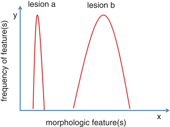
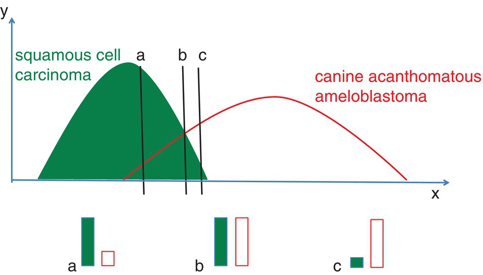
Bell curves can be constructed as simple, two‐dimensional metaphors representing the diversity of morphologic types found within a particular type of lesion (Figure 1.1). For such curves, the diversity of a particular morphologic feature or collection of features within a lesion can be represented along the x‐axis, while the frequency of occurrence of those features in a population of lesions is mapped along the y‐axis. In such a system, a steep and narrow bell curve suggests that relatively little morphologic diversity exists within the lesion type, while a broad‐based bell curve suggests the opposite. Superimposition of these curves graphically demonstrates this concept of overlapping morphologic features (Figure 1.2). Structural overlap between lesions presents a diagnostic challenge for the pathologist and is a concept that will be returned to throughout this book.

Figure 1.1 Bell curves representing lesion diversity and frequency. For a given lesion, the x‐axis can represent a single morphologic feature or set of morphologic features that collectively comprise the lesion in question. The y‐axis represents how common the particular morphologic feature(s) is/are within a group of similar lesions; lesions with a broad curve are morphologically diverse and therefore more difficult to diagnose.

Figure 1.2 Superimposed bell curves are a metaphor for the morphologic overlap between related lesions. Some lesions, like squamous cell carcinoma (SCC) and canine acanthomatous ameloblastoma (CAA) can either be morphologically distinct lesions (extreme right and left edges of the two bell curves) or share multiple features (within the region of curve overlap). Sections a, b, and c represent lesions that are most likely to be SCC, equally likely to be SCC or CAA, or more likely to be CAA, respectively.
It is the opinion of the authors that the examination of histologic features frequently allows the designation of a principal diagnosis along with one or more differential diagnoses. These differential diagnoses are important and should be included in the report sent to the submitting clinician. Assigning a principal diagnosis and accompanying set of differential diagnoses effectively conveys a measure of ambiguity, which may have great value for the clinician. For these reasons, histologically related lesions (differential diagnoses) have been included for each lesion type described in this book.
To assist in this difficult but ultimately rewarding endeavor, the judicious use of appropriate immunohistochemical assays and/or special stains can be invaluable to inform the final diagnosis. Perhaps even more importantly, clinical data, most typically available through the submitting clinician, should be sought out. Patient signalment, anatomic location, and lesion natural history can be invaluable facets of the final diagnosis. Radiographic imaging studies and/or three‐dimensional imaging studies like computed tomography may be available. The opinion of the clinician/ radiologist regarding such studies, or better yet, the diagnostic images themselves, should be reviewed by the pathologist in conjunction with the gross and histological features of the submitted sample.
If not openly offered, the opinion of the submitting clinician should be sought out, as an astute clinician will often have made a preliminary clinical diagnosis prior to submission. This clinical diagnosis may be correct, based upon the clinician's experience, the anatomic location, results of diagnostic imaging studies, signalment of patient, clinical signs, and prior biopsy results. The diagnosis of relatively common oral lesions such as odontogenic cysts and equine cementomas (nodular hypercementosis) are highly dependent upon their anatomic relationship with teeth, jawbones, and/or the paranasal sinuses. Some clinicians have a curious policy of withholding such information from the pathologist in a dubious attempt to “not influence the diagnostic process.” It is likely that these same clinicians would be at a loss if their own clients withheld important clinical information for the same reason.
There is also value in seeking out the opinions of colleagues or even trainees. At academic institutions, such opinions are typically readily available, and such advice may even be offered without asking for it! Useful discussions can also occur in the setting of private diagnostic labs, even in those laboratories staffed by a single pathologist. The common use of digital images facilitates rapid communication, and networks of colleagues around the world are often willing to lend a hand. Finally, following a challenging lesion down the road can be a valuable learning experience in itself. Does the eventual clinical outcome fit the diagnosis, and most importantly, can one learn from it?