

Orthodontic and Surgical Management of Impacted Teeth
Orthodontic and Surgical Management of
IMPACTED
TEETH

Vincent G. Kokich, DDS, MSD
Affiliate Professor
Department of Orthodontics
University of Washington School of Dentistry
Seattle, Washington
David P. Mathews, DDS
Affiliate Associate Professor
Department of Periodontics
University of Washington School of Dentistry
Seattle, Washington
Private practice
Tacoma, Washington

Library of Congress Cataloging-in-Publication Data
Kokich, Vincent G., author.
Orthodontic and surgical management of impacted teeth / Vincent G. Kokich Sr, David P. Mathews.
p. ; cm.
Includes bibliographical references.
ISBN 978-0-86715-445-0 (softcover)
I. Mathews, David P., author. II. Title.
[DNLM: 1. Tooth, Impacted--surgery. 2. Oral Surgical Procedures--methods. 3. Orthodontics-methods. WU 600]
RK521
617.6’43--dc23
2013039998

© 2014 Quintessence Publishing Co, Inc
Quintessence Publishing Co, Inc
4350 Chandler Drive
Hanover Park, IL 60133
www.quintpub.com
5 4 3 2 1
All rights reserved. This book or any part thereof may not be reproduced, stored in a retrieval system, or transmitted in any form or by any means, electronic, mechanical, photocopying, or otherwise, without prior written permission of the publisher.
Editor: Leah Huffman
Design: Ted Pereda
Production: Angelina Sanchez
Printed in China
Contents
Preface
1 Impacted Maxillary Central Incisors
2 Labially Impacted Maxillary Canines
3 Palatally Impacted Canines
4 Impacted Mandibular Canines
5 Impacted Premolars
6 Impacted Mandibular Molars
7 Complications and Adverse Sequelae
Preface
This publication is the culmination of Vince’s vision to write a book chronicling our 39 years of working together in an interdisciplinary manner, treating patients with impacted teeth. There has been very little written on this subject. Vince was the premier educator in orthodontics, and he felt very strongly that we should compile these cases in a very well-crafted book.
Sadly, he passed away before we were able to finish the last chapter. His longtime friend and colleague Dr Peter Shapiro; his son, Dr Vince Kokich, Jr; his daughter, Mary; and his wife, Marilyn, graciously helped me put the final touches on this fine work.
This book includes a chapter on every type of impaction an orthodontist and surgeon will encounter. It describes, in detail, the surgeries to uncover these impactions and the appropriate orthodontic mechanics to move these teeth once they are uncovered. It is the most complete, organized, and up-to-date book on this subject. It is ideal for orthodontists, pedodontists, periodontists, oral and maxillofacial surgeons, and general dentists who treat these types of cases.
We are fortunate to have so many fine orthodontists in Pierce County, Washington, who contributed to some of the cases in this book. They were all students of Vince’s, and we are indebted to them for helping us leave this exceptional legacy.
Vince was the quintessential orthodontist/teacher in the world, and it is only fitting that Quintessence helped us bring this one of a kind book to fruition.
With heartfelt gratitude,
David P. Mathews, DDS

Etiology
The most commonly impacted teeth are the mandibular third molars, followed by the maxillary canines, mandibular second premolars, and maxillary central incisors.1 Several causes of maxillary central incisor impaction have been documented, including crowding, 2 trauma,3,4 root dilaceration,5–9 presence of an odontoma,10,11 presence of a dentigerous cyst,12 or presence of a supernumerary tooth or mesiodens.13–16 Some mesiodentes are located in the maxillary midline, between the two central incisors. In this situation, the supernumerary tooth is not blocking the path of eruption of the maxillary central incisors, and all anterior teeth potentially could erupt normally, including the mesiodens. The solution for this problem is to extract the mesiodens and close the space between the maxillary central incisors orthodontically. However, in most situations, when a mesiodens is present, it is positioned lateral to the maxillary midline and blocks the path of eruption of either the right or left central incisor. If multiple supernumerary teeth are present, both central incisors could become impacted (Fig 1-1).
Fig 1-1 Multiple supernumerary teeth.
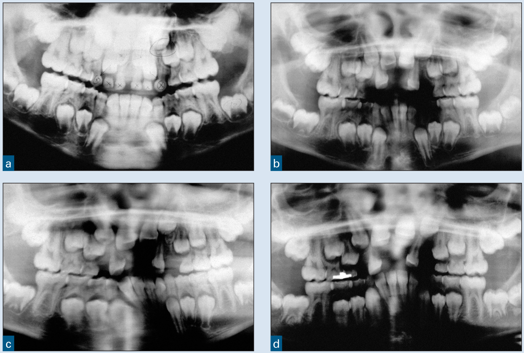
(a) This young patient had two supernumerary teeth that developed coronal to the maxillary central incisors. (b) The supernumerary teeth as well as all six maxillary anterior primary teeth were extracted to facilitate the eruption of the permanent maxillary incisors. (c and d) However, the follicles of the central incisors were damaged during the surgery, and, as a result, the lateral incisors erupted while the central incisors remained impacted.
If the supernumerary tooth or teeth are discovered early and extracted,17 the central incisor could erupt spontaneously (see Fig 1-2). However, the method of extraction and extent of damage to the surrounding area could disrupt the natural eruption of the central incisor(s) and result in impaction. A series of articles published by Marks et al18–22 emphasizes the importance of the integrity of the dental follicle during the normal eruptive process. In their experiments, the researchers intentionally and selectively damaged various parts of the developing tooth bud in an attempt to disrupt the eruptive process and cause impaction of the tooth. However, intentional injury to the root and various parts of the crown of the unerupted tooth did not prevent tooth eruption. But when the dental follicle was damaged or removed, eruption stopped. These researchers believe that the integrity of the dental follicle is critical to the normal eruption of any tooth.
Therefore, if the mesiodens or supernumerary tooth can be extracted without damaging the follicle of the subjacent central incisor, the impediment to eruption will be eradicated, and the impacted central incisor should erupt. This process is illustrated in Fig 1-2. This patient was 8 years old, and the maxillary left central incisor and both maxillary lateral incisors had erupted (see Fig 1-2a). However, the right central incisor had not erupted. A periapical radiograph revealed a supernumerary tooth that was apparently blocking the eruptive path of the right central incisor (see Fig 1-2b). A labial flap was elevated, and the supernumerary tooth was identified and removed without extracting the primary central incisor or damaging the follicle of the developing right central incisor. The flap was repositioned, and the teeth were allowed to erupt. After 1 year, the right central incisor had erupted spontaneously without orthodontic intervention (see Fig 1-2c). Therefore, if the supernumerary tooth is diagnosed early and removed surgically without compromising the follicle of the impacted central incisor, the submerged tooth should erupt spontaneously.
Fig 1-2 Interceptive removal of a supernumerary tooth.
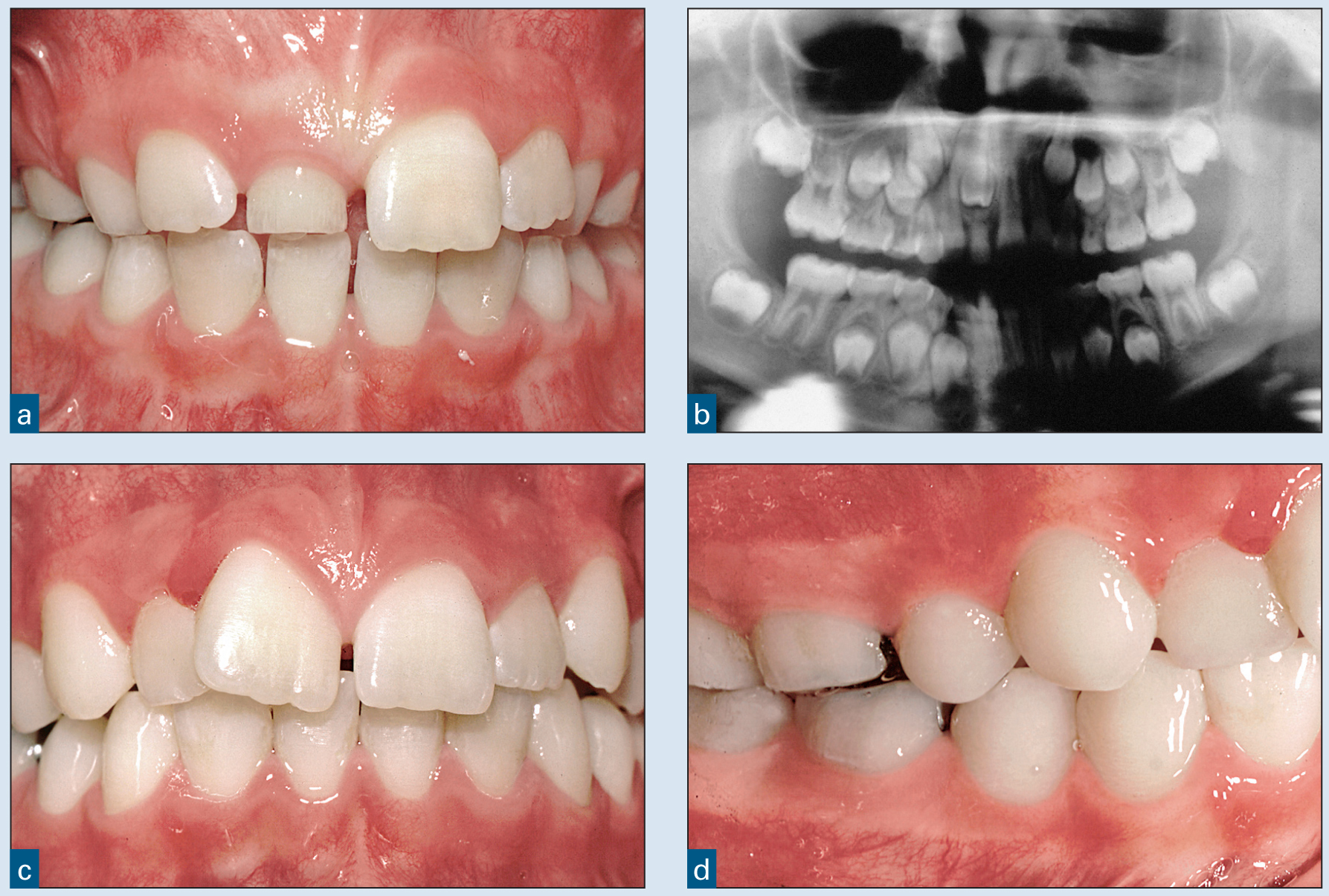
(a) This patient was 7 years, 8 months old. Her maxillary right central incisor was impacted within the alveolus. (b) The panoramic radiograph showed that a supernumerary tooth was blocking the path of eruption of the central incisor. The supernumerary tooth was removed without damaging the follicle of the right central incisor. (c) Eventually, the maxillary right central incisor erupted spontaneously without any further intervention. (d) This patient’s occlusion was developing normally, and the parents chose not to proceed with any further orthodontic treatment.
However, in some situations, the proximity of the supernumerary tooth and the central incisor is so close that damage to the follicle of the impacted central incisor is unavoidable and inevitable. In other situations, an odontoma or dentigerous cyst may be present and in close proximity to the central incisor crown. Removal of the odontoma or cyst will usually damage the central incisor follicle, and the tooth will stop erupting. Finally, in some situations, a severely dilacerated root will cause impaction of the central incisor as a result of diversion of the tooth’s eruptive path, placing it in an oblique position within the alveolus (see Fig 1-9). In all of these situations, it is best to uncover the impacted central incisor at the same time that the other surgery is being done, assuming the root of the central incisor is adequately developed.
Preoperative Orthodontics
Usually, an impacted central incisor is recognized during the mixed dentition. At that time, all maxillary and mandibular central and lateral incisors are erupted, except for the impacted central incisor. The first step to facilitating spontaneous eruption is to extract any supernumerary teeth. If the tooth does not erupt autonomously, orthodontic treatment must be initiated to erupt the tooth.
Brackets should be placed on the remaining central incisor and the two maxillary lateral incisors. This bracket placement typically provides sufficient anchorage to erupt the impacted tooth. If the contralateral central incisor and adjacent lateral incisors are tipped toward one another, the space is opened using a compressed-coil spring. In this situation, it is necessary to place bands or brackets on the permanent maxillary first molars to provide anchorage during the course of orthodontic treatment.
After sufficient space has been established, a rectangular stabilizing wire is placed in the maxillary brackets. A loop may be placed in the archwire to temporarily anchor the attachment that will be placed on the impacted central incisor during the uncovering procedure. At this point, the patient is referred to the surgeon to uncover the impacted central incisor.
Methods of Uncovering
There are four methods for uncovering an impacted maxillary central incisor: simple excision of tissue (gingivectomy), apically positioned flap (APF), the closed eruption technique, and surgical replantation. Central incisors are usually impacted labially. The apical location of the incisor will dictate the type of uncovering technique. Choosing the correct method will create the most stable and esthetic outcome after orthodontic eruption of the impacted tooth.
Most labially impacted central incisors are uncovered with the closed eruption technique. 23 Occasionally, the central incisor is located near or slightly coronal to the adjacent cementoenamel junction. If there is a wide zone of attached gingiva, a gingivectomy can be performed. The authors do not recommend an APF for uncovering maxillary central incisors because of the stability and esthetic problems associated with this technique.
Gingivectomy
Gingivectomy can be performed if the resultant uncovering leaves at least 3 mm of gingiva around the exposed tooth. The gingivectomy should remove approximately two-thirds of the tissue covering the crown of the impacted tooth. A bracket and/or dressing can be placed to ensure that the tissue does not re-cover the tooth. There are few indications for this method of uncovering impacted maxillary central incisors. In most situations, the impacted tooth will be located at or above the mucogingival junction (Fig 1-3). If the tooth were at this level and an excisional approach were used, it would remove most of the gingiva, leaving inadequate attached gingiva over the labial portion of the crown (see Fig 1-3).
Fig 1-3 Excisional uncovering (gingivectomy) of a maxillary left central incisor.
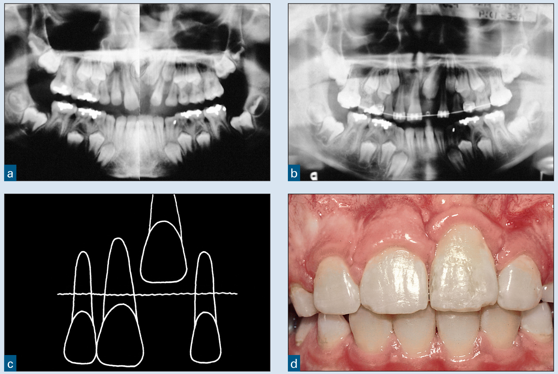
(a) The maxillary left central incisor in this 8½-year-old boy had not erupted. His occlusion was developing normally, but the right central incisor had drifted mesially. (b) Orthodontic alignment was initiated to create space for the impacted central incisor, but after 6 months, the tooth had not erupted. (c) A diagram shows that the incisal edge of the impacted central incisor was above the mucogingival junction. A gingivectomy was used to uncover the tooth, and it was erupted orthodontically into position. (d) After this phase of orthodontics, the gingival margin of the left central incisor was located apical to that of the right central incisor. In addition, the gingival margin of the left central incisor was thickened, rolled, and unesthetic.
Apically positioned flap
A second type of surgical procedure for uncovering impacted maxillary central incisors is an APF (Figs 1-4 and 1-5). This procedure will create a predictable zone of attached gingiva, because it apically positions labial gingiva over the impacted central incisor. However, there are two problems associated with this technique: reintrusion and esthetics.24 Therefore, the authors no longer use this technique to uncover maxillary central incisors.
Fig 1-4 APF involving a horizontally impacted maxillary central incisor.
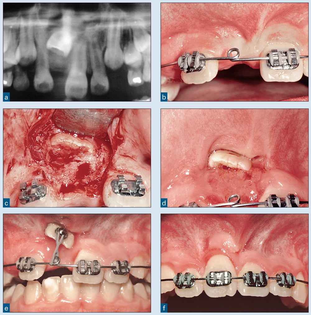
(a) The pretreatment radiograph shows the position of the impacted right central incisor, rotated 90 degrees. (b) Brackets were placed on the adjacent teeth, and sufficient space was created for the right central incisor. (c) An APF was reflected, bone was removed to uncover the crown, and the flap was sutured apically to leave the tooth uncovered. (d to f) Three weeks after uncovering, an elastomeric chain was used to erupt the right central incisor, and brackets were placed to position the crown and root of the formerly impacted central incisor.
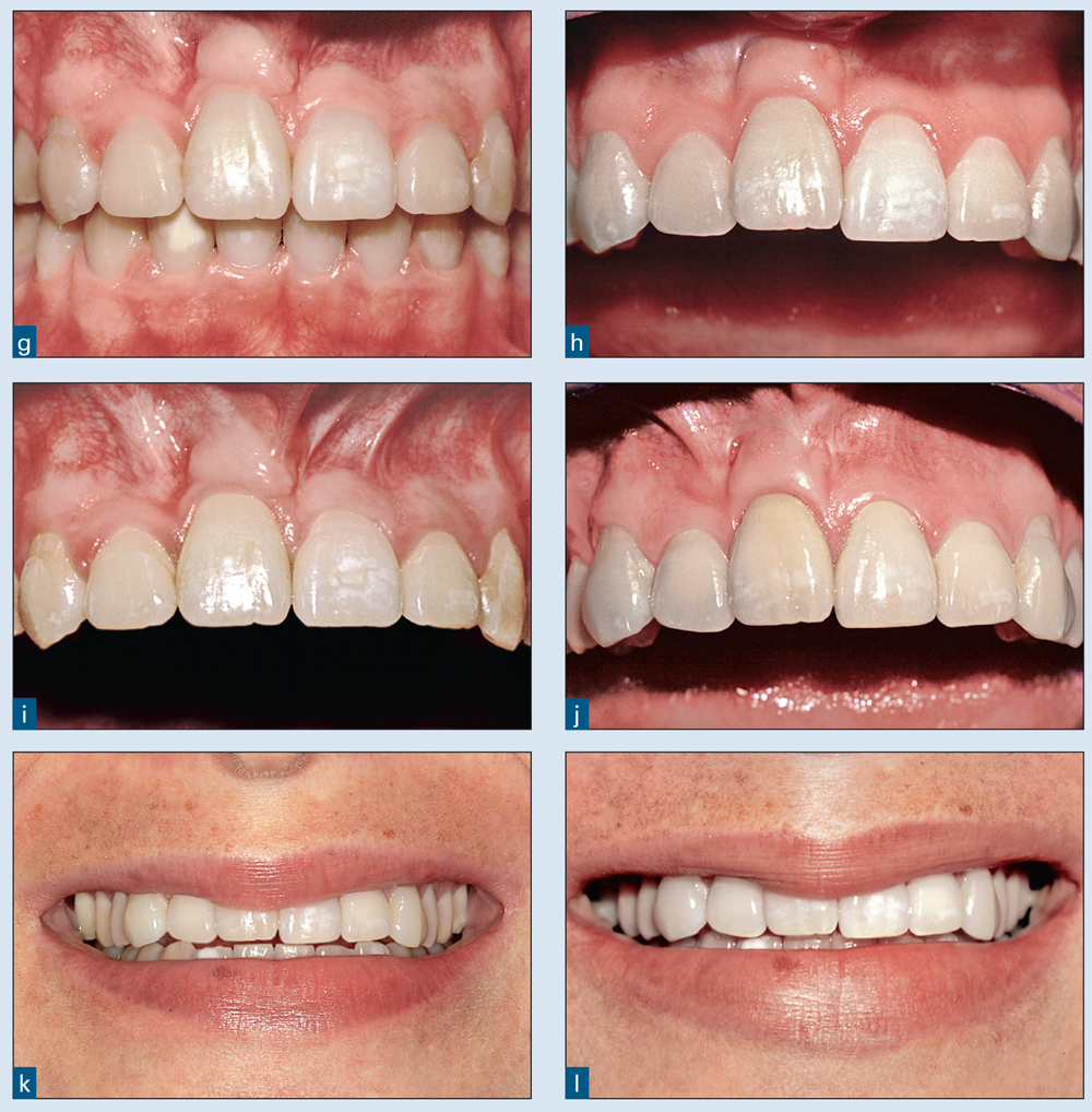
(g) After completion of the orthodontic treatment, the gingival margin of the right central incisor was more apical than that of the left central incisor. In addition, the gingiva was considerably thicker and unesthetic. (h) Five years after orthodontic treatment, the maxillary right central incisor had reintruded. This relapse was probably due to the pull of the mucosa (pseudofrena). (i) The tooth was retreated to level the incisal edges. (j) Twenty-five years later, the right central incisor has reintruded, and the gingival margins are uneven again. (k and l) Five and 25 years after orthodontics, respectively, the incisal edge discrepancies are apparent when the patient smiles.
Fig 1-5 APF to uncover two labially impacted central incisors.
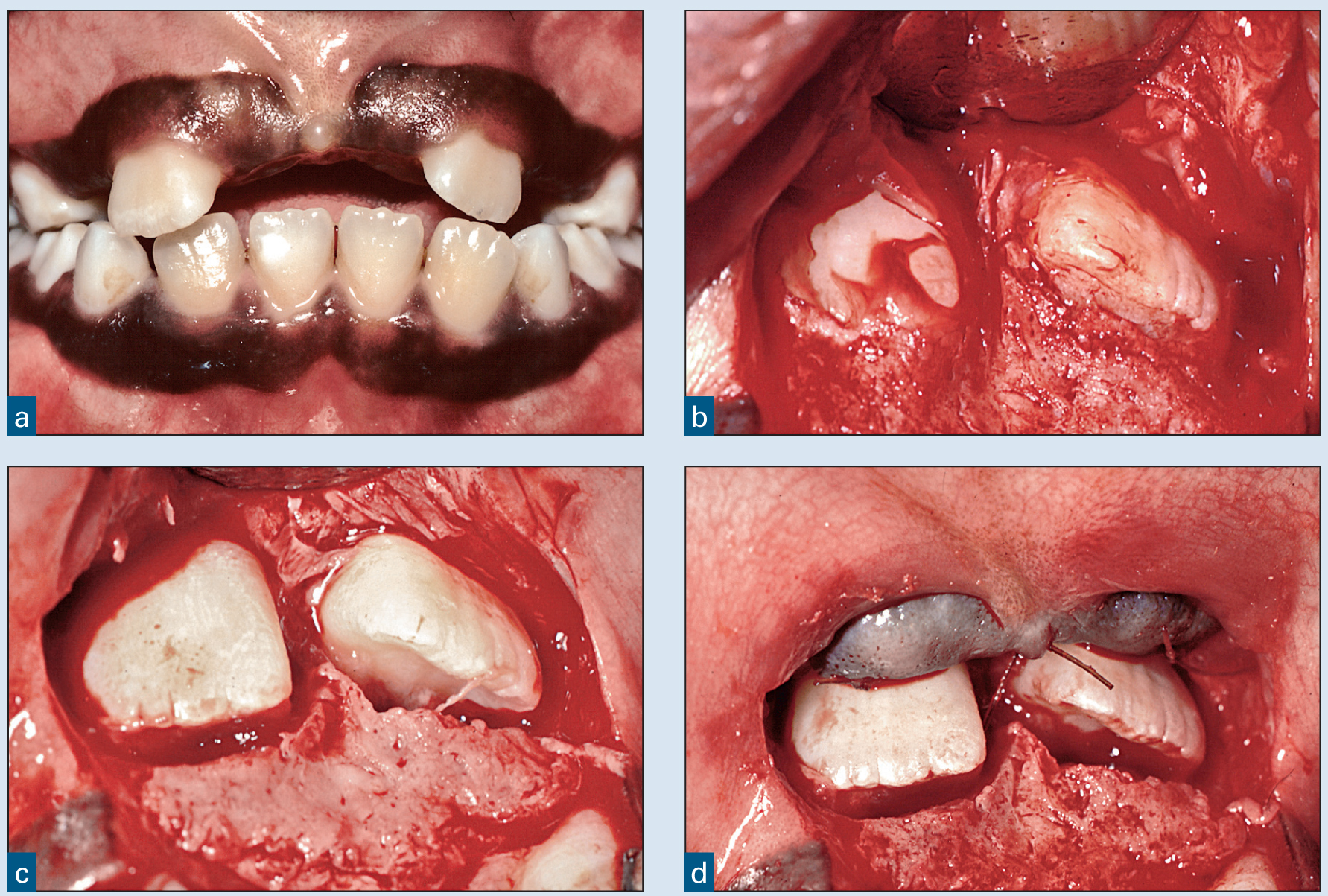
(a) One year after removal of supernumerary teeth, the permanent central incisors are still impacted. (b) A pedicle flap was reflected using midcrestal and vertical incisions, exposing the impacted central incisors. (c and d) Bone was removed to uncover the crowns of the two incisors, and the flap was apically positioned, leaving the incisors uncovered.
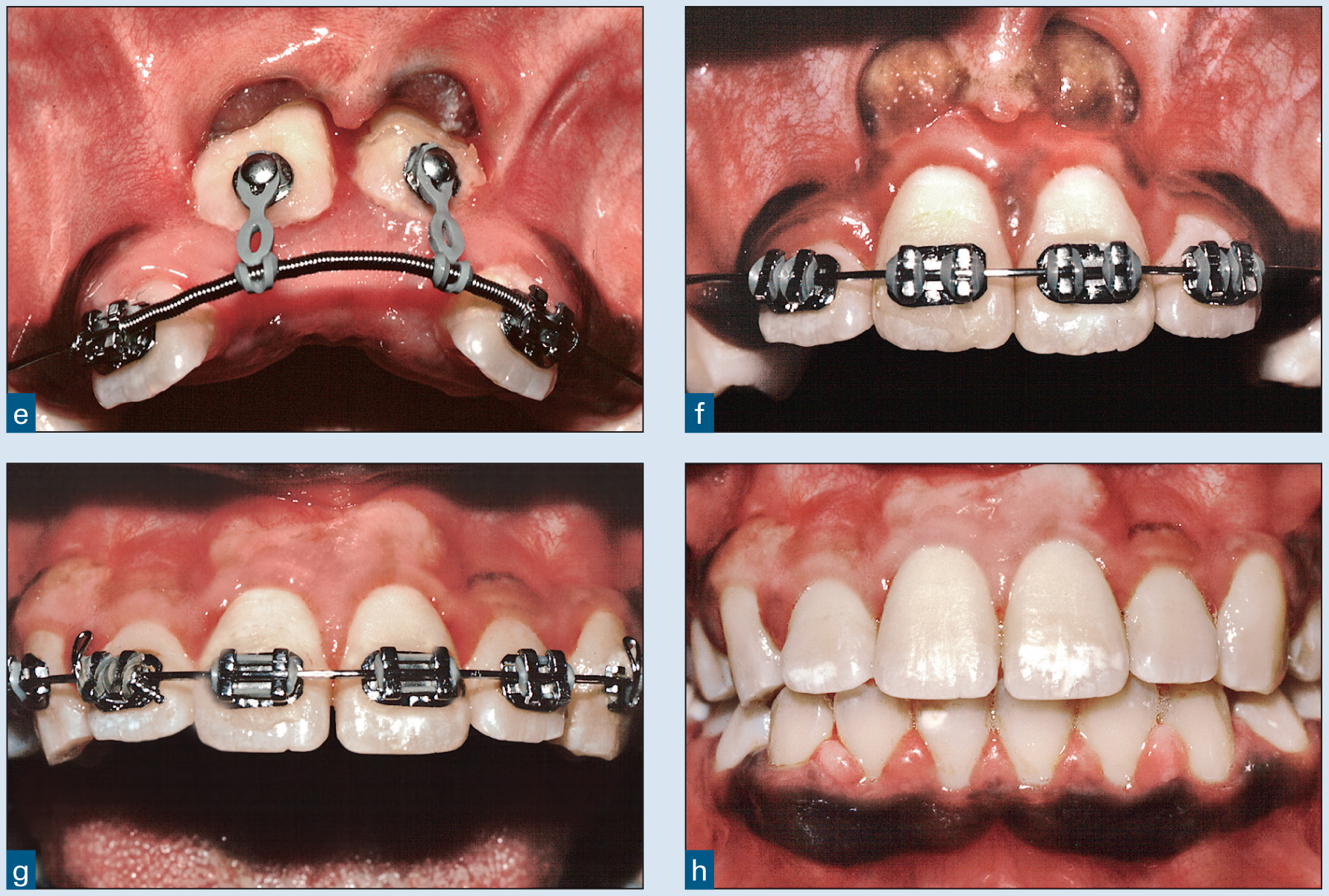
(e) Six weeks later, elastomeric chains were attached, and the eruption process was initiated. (f) One year later, the teeth were in ideal position. Note the bulky, apically displaced gingival tissue and disruption of the mucogingival junction. (g) Gingivoplasty was performed to eliminate the bulky tissue. Note the alteration of the natural pigmentation. (h) Two years later, the teeth have remained stable.
If an APF were used to uncover a complex high labial impaction, it would be likely that gingival margin discrepancies would occur. This consequence is illustrated in Fig 1-4. The maxillary right central incisor has been moved into its proper location, but the gingival margin is more apical than that of the adjacent central incisor (see Figs 1-4f and 1-4g). This case illustrates one of the reasons why the closed eruption technique is used to uncover most labially impacted teeth: It maintains the original gingival architecture. An exception is the ectopic labially impacted tooth, which must remain uncovered after surgery. This situation requires an APF so that appropriate orthodontic mechanics can be applied to properly position the tooth.
Closed eruption technique
The third technique for uncovering an impacted maxillary central incisor is the closed eruption technique.23–27 This technique involves reflecting a flap, uncovering the impacted tooth, placing an attachment, repositioning the flap, and erupting the tooth through the crest of the alveolar ridge (Figs 1-6 and 1-7). This technique results in the most natural-appearing gingiva over the erupted tooth.25,28,29 The crown length is usually commensurate with the nonimpacted contralateral maxillary central incisor, resulting in a more esthetic result. The closed eruption technique also eliminates the reintrusion problem after the tooth has been erupted.
The closed eruption technique is illustrated in Fig 1-6. This patient had a supernumerary tooth coronal to the maxillary left central incisor (see Fig 1-6a). Brackets were placed on the erupted maxillary incisors, and space was apportioned for the impacted central incisor (see Figs 1-6b and 1-6c). A midcrestal incision was made in the edentulous ridge and joined with vertical incisions (see Fig 1-6d), and a full-thickness pedicle flap was reflected from the edentulous ridge. The supernumerary tooth was then located (see Fig 1-6e), but it communicated with the follicle of the impacted central incisor. The follicle was perforated when the supernumerary tooth was extracted (see Fig 1-6f). Because the follicle was disrupted, the central incisor would not erupt spontaneously, and therefore the impacted tooth was uncovered. There is usually a thin shell of bone covering part of the tooth. Approximately two-thirds of the crown was exposed with appropriate bone removal using curettes and surgical round burs. The area was isolated with hemostatic agents such as Surgicel (Ethicon) or Hemodent (Premier USA) cotton pledgets. The tooth was etched, and a bonding agent was placed. It is imperative that the field be kept clean and dry. With experience, hemostasis and a dry field can be accomplished in even the most difficult areas to access.
Fig 1-6 Closed eruption technique.
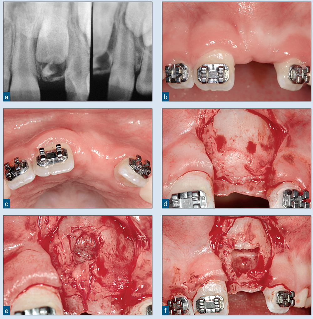
(a) The radiograph shows a supernumerary tooth and a labially impacted central incisor. (b and c) Brackets were placed, and appropriate space was created to allow eruption of the left central incisor. (d) A pedicle flap was reflected from the crest of the ridge. The supernumerary tooth and left central incisor were impacted within the alveolus and not visible. (e) A curette was used to remove the thin labial bone to find the supernumerary tooth. (f) The supernumerary tooth was removed, and the central incisor was now visible.
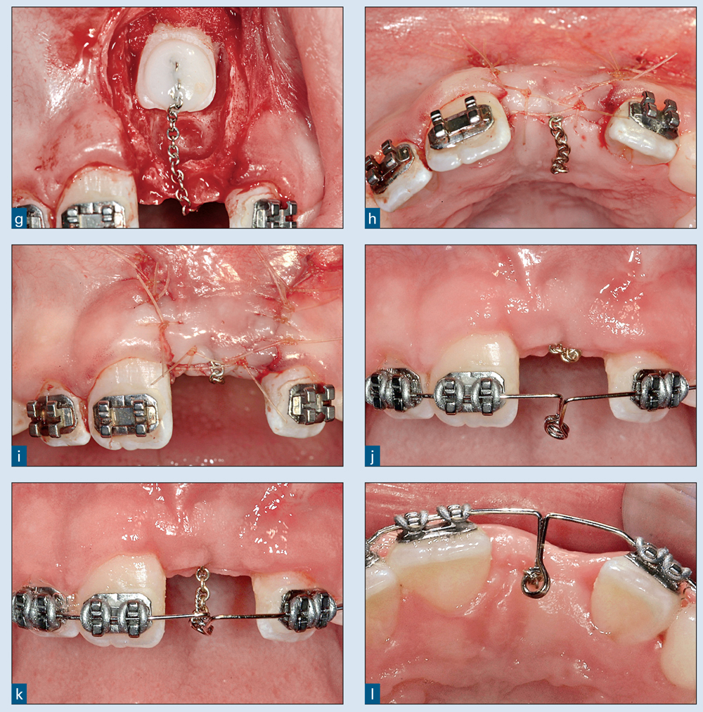
(g) Bone was removed from the crown of the central incisor, and a gold chain was bonded directly to the labial surface of the tooth. (h and i) The flap was repositioned and sutured. The chain exited through the incision at the midcrestal area. (j) Six weeks later, a Ballista spring was placed and was in the inactive state (ie, unattached). (k) The Ballista spring was activated and ligated to the gold chain. (l) The occlusal view of the activated spring demonstrates the midcrestal force.
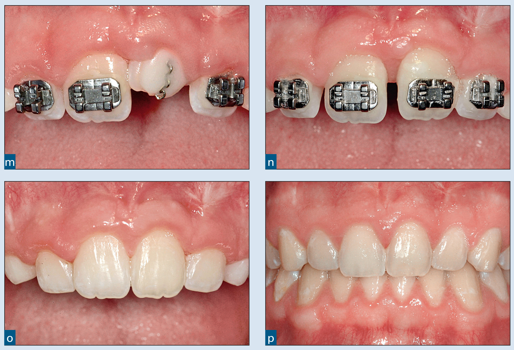
(m) The central incisor erupted through the crest of the ridge. The gingival margin was even with that of the right central incisor. (n) A bracket was placed for orthodontic finishing. (o) At completion, the tooth had a normal mucogingival junction and no pseudofrena. (p) Five years later, the gingival margins of the two central incisors are even and reintrusion has not occurred. (Orthodontics courtesy of Dr Vince Kokich, Jr, Tacoma, Washington.)
Once the bonding agent is placed and cured, the bonding process can continue. If the area gets contaminated with blood or debris after placement of the bonding agent, it can be cleaned with an alcohol wipe. At this point, a small cleat can be bonded to the tooth, and a chain can be ligated to the cleat. The chain can also be bonded directly to the tooth (see Fig 1-6g). The chain should have small enough links so the orthodontist can clip and remove one or two links as the tooth erupts. The chain should be malleable enough to avoid breakage but hard enough to avoid stretching as force is applied. Prefabricated chains with brackets for specific teeth are available (GAC International) and are easy to bond to impacted teeth. Fourteen-carat gold replacement chain with appropriate link size can also be effective. This can be purchased from a jeweler or pawnshop and is relatively inexpensive.
The pedicle flap is then returned to its original position and sutured. The chain is covered by the flap and exits at the midcrestal incision (see Fig 1-6h). The gold chain is ligated to the bracket on the adjacent tooth. A small ligating wire or an elastomeric ligature works well to fasten the chain to the bracket. The orthodontist may begin eruption of the tooth within 1 to 2 weeks.
The closed eruption technique can also be used when the tooth is impacted high in the vestibule on the labial aspect (Fig 1-8). In Fig 1-9, the maxillary right central incisor was impacted high in the vestibule near the base of the nasal spine. It was also positioned horizontally, which further complicated the surgical access (see Figs 1-9a and 1-9b). Initially, the maxillary incisors were banded (see Fig 1-9c) and the teeth were aligned, leaving a space for the impacted right central incisor (see Figs 1-9d and 1-9e). The patient was then ready to have the tooth uncovered. The use of an APF in this situation would be injudicious, so the authors chose the closed eruption technique.
Fig 1-7 Closed eruption technique involving a labially impacted maxillary central incisor.
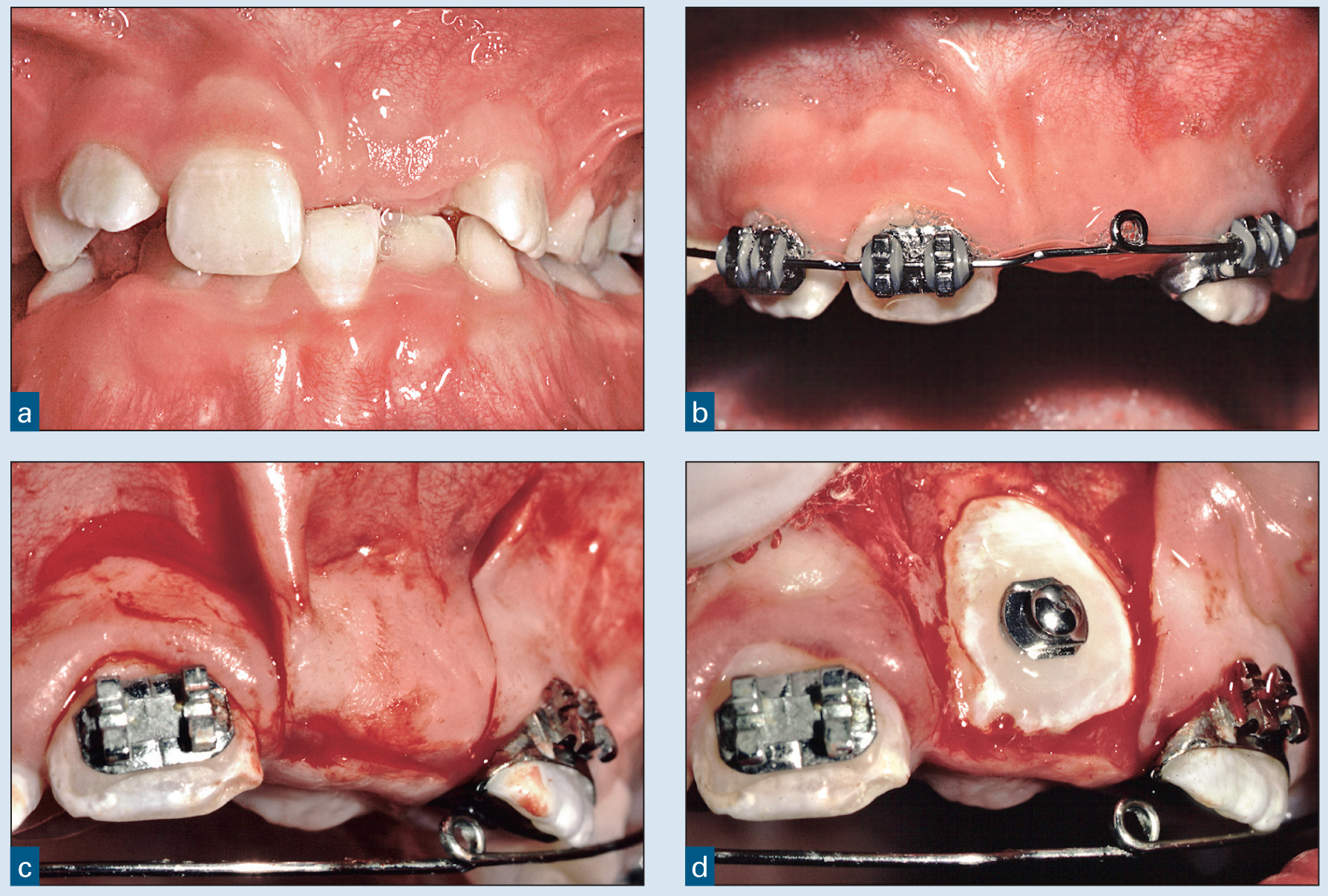
(a) This 10-year-old patient’s left central incisor had not erupted. (b) Orthodontic treatment was initiated to create sufficient space between the right central and left lateral incisors. (c and d) A pedicle flap was reflected from the crest of the ridge, and a button was bonded to the labial of the impacted central incisor.
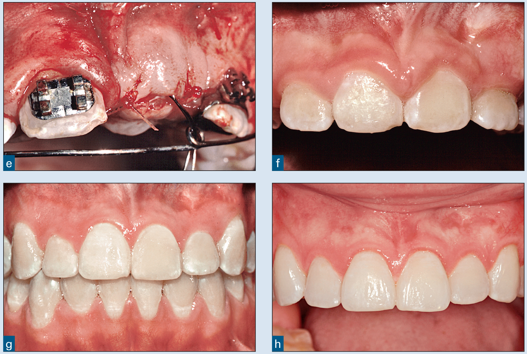
(e) The flap was repositioned and sutured, and elastomeric chains were used to erupt the tooth. (f) The central incisor was erupted orthodontically through the crest of the ridge. The gingival margins were at a commensurate level. (g) (h)